3
views
0
recommends
+1 Recommend
1 collections
    0
    shares
      • Record: found
      • Abstract: found
      • Article: found
      Is Open Access

      Host protein kinases required for SARS-CoV-2 nucleocapsid phosphorylation and viral replication

      research-article
      1 , 2 , 3 , 4 , 5 , 6 , § , 7 , 1 , 2 , 8 , 1 , 2 , 1 , 2 , 3 , 1 , 2 , 9 , 1 , 2 , 3 , 1 , 2 , 1 , 20 , 1 , 2 , 1 , 2 , 1 , 2 , 9 , 1 , 2 , 10 , 4 , 11 , 3 , 4 , 3 , 4 , 1 , 12 , 8 , 13 , 6 , 6 , 6 , 6 , 6 , 14 , 6 , 14 , 6 , 7 , 7 , 7 , 7 , 15 , 15 , 15 , 16 , 17 , 18 , 19 , 18 , 18 , 8 , § , 1 , 2 , 20 , 21 , § , 1 , 22 , 23 , § , 6 , 24 , 25 , §
      Science signaling

      Read this article at

      Bookmark
          There is no author summary for this article yet. Authors can add summaries to their articles on ScienceOpen to make them more accessible to a non-specialist audience.

          Abstract

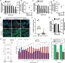
          Multiple coronaviruses have emerged independently in the past 20 years that cause lethal human diseases. Although vaccine development targeting these viruses has been accelerated substantially, there remain patients requiring treatment who cannot be vaccinated or who experience breakthrough infections. Understanding the common host factors necessary for the life cycles of coronaviruses may reveal conserved therapeutic targets. Here, we used the known substrate specificities of mammalian protein kinases to deconvolute the sequence of phosphorylation events mediated by three host protein kinase families (SRPK, GSK-3, and CK1) that coordinately phosphorylated a cluster of serine and threonine residues in the viral N protein, which is required for viral replication. We also showed that loss or inhibition of SRPK1/2, which we propose initiates the N protein phosphorylation cascade, compromised the viral replication cycle. Because these phosphorylation sites are highly conserved across coronaviruses, inhibitors of these protein kinases may not only have therapeutic potential against COVID-19, but also may be broadly useful against multiple coronavirus-mediated diseases.

          Related collections

          Most cited references89

          • Record: found
          • Abstract: found
          • Article: found
          Is Open Access

          limma powers differential expression analyses for RNA-sequencing and microarray studies

          limma is an R/Bioconductor software package that provides an integrated solution for analysing data from gene expression experiments. It contains rich features for handling complex experimental designs and for information borrowing to overcome the problem of small sample sizes. Over the past decade, limma has been a popular choice for gene discovery through differential expression analyses of microarray and high-throughput PCR data. The package contains particularly strong facilities for reading, normalizing and exploring such data. Recently, the capabilities of limma have been significantly expanded in two important directions. First, the package can now perform both differential expression and differential splicing analyses of RNA sequencing (RNA-seq) data. All the downstream analysis tools previously restricted to microarray data are now available for RNA-seq as well. These capabilities allow users to analyse both RNA-seq and microarray data with very similar pipelines. Second, the package is now able to go past the traditional gene-wise expression analyses in a variety of ways, analysing expression profiles in terms of co-regulated sets of genes or in terms of higher-order expression signatures. This provides enhanced possibilities for biological interpretation of gene expression differences. This article reviews the philosophy and design of the limma package, summarizing both new and historical features, with an emphasis on recent enhancements and features that have not been previously described.
            Bookmark
            • Record: found
            • Abstract: found
            • Article: not found

            Early Transmission Dynamics in Wuhan, China, of Novel Coronavirus–Infected Pneumonia

            Abstract Background The initial cases of novel coronavirus (2019-nCoV)–infected pneumonia (NCIP) occurred in Wuhan, Hubei Province, China, in December 2019 and January 2020. We analyzed data on the first 425 confirmed cases in Wuhan to determine the epidemiologic characteristics of NCIP. Methods We collected information on demographic characteristics, exposure history, and illness timelines of laboratory-confirmed cases of NCIP that had been reported by January 22, 2020. We described characteristics of the cases and estimated the key epidemiologic time-delay distributions. In the early period of exponential growth, we estimated the epidemic doubling time and the basic reproductive number. Results Among the first 425 patients with confirmed NCIP, the median age was 59 years and 56% were male. The majority of cases (55%) with onset before January 1, 2020, were linked to the Huanan Seafood Wholesale Market, as compared with 8.6% of the subsequent cases. The mean incubation period was 5.2 days (95% confidence interval [CI], 4.1 to 7.0), with the 95th percentile of the distribution at 12.5 days. In its early stages, the epidemic doubled in size every 7.4 days. With a mean serial interval of 7.5 days (95% CI, 5.3 to 19), the basic reproductive number was estimated to be 2.2 (95% CI, 1.4 to 3.9). Conclusions On the basis of this information, there is evidence that human-to-human transmission has occurred among close contacts since the middle of December 2019. Considerable efforts to reduce transmission will be required to control outbreaks if similar dynamics apply elsewhere. Measures to prevent or reduce transmission should be implemented in populations at risk. (Funded by the Ministry of Science and Technology of China and others.)
              Bookmark
              • Record: found
              • Abstract: found
              • Article: not found

              Imbalanced Host Response to SARS-CoV-2 Drives Development of COVID-19

              Summary Viral pandemics, such as the one caused by SARS-CoV-2, pose an imminent threat to humanity. Because of its recent emergence, there is a paucity of information regarding viral behavior and host response following SARS-CoV-2 infection. Here we offer an in-depth analysis of the transcriptional response to SARS-CoV-2 compared with other respiratory viruses. Cell and animal models of SARS-CoV-2 infection, in addition to transcriptional and serum profiling of COVID-19 patients, consistently revealed a unique and inappropriate inflammatory response. This response is defined by low levels of type I and III interferons juxtaposed to elevated chemokines and high expression of IL-6. We propose that reduced innate antiviral defenses coupled with exuberant inflammatory cytokine production are the defining and driving features of COVID-19.
                Bookmark

                Author and article information

                Journal
                101465400
                34171
                Sci Signal
                Sci Signal
                Science signaling
                1945-0877
                1937-9145
                5 January 2023
                25 October 2022
                25 October 2022
                10 January 2023
                : 15
                : 757
                : eabm0808
                Affiliations
                [1 ]Meyer Cancer Center, Weill Cornell Medicine, New York, NY 10021, USA.
                [2 ]Department of Medicine, Weill Cornell Medicine, New York, NY 10021, USA.
                [3 ]Englander Institute for Precision Medicine, Institute for Computational Biomedicine, Weill Cornell Medicine, New York, NY 10021, USA.
                [4 ]Department of Physiology and Biophysics, Weill Cornell Medicine, New York, NY 10065, USA.
                [5 ]Tri-Institutional PhD Program in Computational Biology & Medicine, Weill Cornell Medicine/Memorial Sloan Kettering Cancer Center/The Rockefeller University, New York, NY 10021, USA.
                [6 ]Department of Molecular Genetics and Microbiology, Duke University School of Medicine, Durham, NC 27710, USA.
                [7 ]Cell Signaling Technology, Danvers, MA 01923, USA.
                [8 ]New York University, Grossman School of Medicine, New York, NY 10016, USA.
                [9 ]Weill Cornell Graduate School of Medical Sciences, Cell and Developmental Biology Program, New York, NY 10065, USA.
                [10 ]Division of Gastroenterology and Hepatology, Department of Medicine, Weill Cornell Medicine, New York, NY 10065, USA.
                [11 ]Department of Surgery, Weill Cornell Medicine, New York, NY 10065, USA.
                [12 ]Department of Dermatology, Weill Cornell Medicine, New York, NY 10065, USA.
                [13 ]Department of Microbiology and Molecular Genetics, Chao Family Comprehensive Cancer Center, University of California Irvine School of Medicine, Irvine, CA 92868, USA.
                [14 ]Department of Cell Biology, Duke University School of Medicine, Durham, NC 27710, USA.
                [15 ]Broad Institute of MIT & Harvard, Cambridge, MA 02142, USA.
                [16 ]Department of Pathology, Harvard Medical School, Boston, MA 02115, USA.
                [17 ]Cancer Center and Department of Pathology, Massachusetts General Hospital, Boston, MA 02114, USA.
                [18 ]School of Cellular and Molecular Medicine, University of Bristol, Bristol, BS8 1TD, UK.
                [19 ]Proteomics Facility, University of Bristol, Bristol, BS8 1TD, UK.
                [20 ]Dana-Farber Cancer Institute, Harvard Medical School, Boston, MA 02215, USA.
                [21 ]Department of Cell Biology, Harvard Medical School, Boston, MA 02115, USA.
                [22 ]Department of Pharmacology, Weill Cornell Medicine, New York, NY 10065, USA.
                [23 ]Department of Biochemistry, Weill Cornell Medicine, New York, NY 10065, USA.
                [24 ]Duke Human Vaccine Institute, Duke University School of Medicine Durham, NC 27710, USA.
                [25 ]Duke Cancer Institute, Duke University School of Medicine, Durham, NC 27710, USA.
                Author notes
                [*]

                These authors contributed equally to this work.

                Author contributions: T.M.Y., B.E.H., L.C.C., and N.S.H. conceived the project, designed experiments, processed data, and wrote the manuscript. T.X.J. and B.E.N.P. performed the experiments shown in Fig. 1. T.M.L., A.P.P., T.M.Y., S.L.T., P.V.H., and S.A.B. performed mass spectrometry analysis and processed the mass spectrometry data. J.L.J., T.L., and K.M.L. performed the experiments shown in Fig. 2. B.E.H., N.S.H., J.D.T., K.N.B., C.E.H., R.R.C., A.T.H., A.T., X.Z., and C.M.S. performed the experiments shown in Figs. 3 and 4 and fig. S4. D.K.B., S.J.V.N., N.K., E.R.K., M.N.M., K.S.G., A.D.D., K.H., M.K.W., and E.P. assisted with experiments. B.M.C., and T.M.Y. performed the evolutionary conservation analysis. T.M.Y. and A.K. performed computational analysis of the kinase library data. T.M.Y., S.K.A., F.A., and G.G. performed computational analysis of the proteomics data. L.H., R.E.S., S.C., H.W., O.E., E.P., G.L., and D.M. participated in discussions. J.B., B.R.t., L.C.C., B.E.H., and N.S.H. supervised the work.

                Article
                NIHMS1851690
                10.1126/scisignal.abm0808
                9830954
                36282911
                e6fffc1e-9b01-4f6e-ac04-5c1dcb78578f

                This work is licensed under a Creative Commons Attribution 4.0 International (CC BY 4.0) license, which permits unrestricted use, distribution, and reproduction in any medium, provided the original work is properly cited. To view a copy of this license, visit https://creativecommons.org/licenses/by/4.0/. This license does not apply to figures/photos/artwork or other content included in the article that is credited to a third party; obtain authorization from the rights holder before using this material.

                History
                Categories
                Article

                Comments

                Comment on this article