49
views
0
recommends
+1 Recommend
0 collections
    0
    shares
      • Record: found
      • Abstract: found
      • Article: found
      Is Open Access

      Imaging fictive locomotor patterns in larval Drosophila

      research-article

      Read this article at

      Bookmark
          There is no author summary for this article yet. Authors can add summaries to their articles on ScienceOpen to make them more accessible to a non-specialist audience.

          Abstract

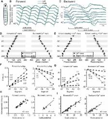
          We have established a preparation in larval Drosophila to monitor fictive locomotion simultaneously across abdominal and thoracic segments of the isolated CNS with genetically encoded Ca 2+ indicators. The Ca 2+ signals closely followed spiking activity measured electrophysiologically in nerve roots. Three motor patterns are analyzed. Two comprise waves of Ca 2+ signals that progress along the longitudinal body axis in a posterior-to-anterior or anterior-to-posterior direction. These waves had statistically indistinguishable intersegmental phase delays compared with segmental contractions during forward and backward crawling behavior, despite being ∼10 times slower. During these waves, motor neurons of the dorsal longitudinal and transverse muscles were active in the same order as the muscle groups are recruited during crawling behavior. A third fictive motor pattern exhibits a left-right asymmetry across segments and bears similarities with turning behavior in intact larvae, occurring equally frequently and involving asymmetry in the same segments. Ablation of the segments in which forward and backward waves of Ca 2+ signals were normally initiated did not eliminate production of Ca 2+ waves. When the brain and subesophageal ganglion (SOG) were removed, the remaining ganglia retained the ability to produce both forward and backward waves of motor activity, although the speed and frequency of waves changed. Bilateral asymmetry of activity was reduced when the brain was removed and abolished when the SOG was removed. This work paves the way to studying the neural and genetic underpinnings of segmentally coordinated motor pattern generation in Drosophila with imaging techniques.

          Related collections

          Most cited references64

          • Record: found
          • Abstract: found
          • Article: not found

          The expression pattern of the Drosophila vesicular glutamate transporter: a marker protein for motoneurons and glutamatergic centers in the brain.

          To determine the functions of genes in distinct tissues during the development of Drosophila, it is often desirable to have genetic tools for targeted gene expression in restricted subsets of cells. Here, we report the identification of the enhancer trap line OK371-Gal4, which is expressed in a defined subset of neurons from embryonic stage 15 to adulthood. In the ventral nerve chord, it is expressed almost exclusively in motoneurons and in the brain in a limited number of neuronal clusters. The OK371 enhancer trap element is inserted in the proximity of the annotated gene CG9887, which encodes a Drosophila vesicular glutamate transporter (DVGLUT). In situ hybridization experiments using antisense probes against the mRNAs of DVGLUT and neighboring genes confirm that OK371-Gal4 detects an enhancer of DVGLUT. DVGLUT-specific antibodies detect its expression in identifiable motoneurons, which are known to be glutamatergic in Drosophila. DVGLUT initially appears in small cytoplasmic punctae in the somata of these motoneurons. As development proceeds, DVGLUT-positive particles are transported along motor axons and become concentrated at neuromuscular junctions (NMJs), where they colocalize with the synaptic vesicle marker synaptotagmin. We find that the DVGLUT-specific antibodies are valuable tools for the identification of motoneurons and other glutamatergic neurons. In addition, the OK371-Gal4 line can be used for the targeted expression of any gene in these cells. Given that vesicular glutamate transporters are essential for the uptake of the neurotransmitter glutamate into synaptic vesicles these tools provide a means to test gene function in these functionally important neurons.
            Bookmark
            • Record: found
            • Abstract: found
            • Article: not found

            Sampling properties of the spectrum and coherency of sequences of action potentials.

            The spectrum and coherency are useful quantities for characterizing the temporal correlations and functional relations within and between point processes. This article begins with a review of these quantities, their interpretation, and how they may be estimated. A discussion of how to assess the statistical significance of features in these measures is included. In addition, new work is presented that builds on the framework established in the review section. This work investigates how the estimates and their error bars are modified by finite sample sizes. Finite sample corrections are derived based on a doubly stochastic inhomogeneous Poisson process model in which the rate functions are drawn from a low-variance gaussian process. It is found that in contrast to continuous processes, the variance of the estimators cannot be reduced by smoothing beyond a scale set by the number of point events in the interval. Alternatively, the degrees of freedom of the estimators can be thought of as bounded from above by the expected number of point events in the interval. Further new work describing and illustrating a method for detecting the presence of a line in a point process spectrum is also presented, corresponding to the detection of a periodic modulation of the underlying rate. This work demonstrates that a known statistical test, applicable to continuous processes, applies with little modification to point process spectra and is of utility in studying a point process driven by a continuous stimulus. Although the material discussed is of general applicability to point processes, attention will be confined to sequences of neuronal action potentials (spike trains), the motivation for this work.
              Bookmark
              • Record: found
              • Abstract: found
              • Article: not found

              Ca2+ buffering and action potential-evoked Ca2+ signaling in dendrites of pyramidal neurons.

              The effect of the fluorescent Ca2+ indicator dye Fura-2 on Ca2+ dynamics was studied in proximal apical dendrites of neocortical layer V and hippocampal CA1 pyramidal neurons in rat brain slices using somatic whole-cell recording and a charge-coupled device camera. A single action potential evoked a transient increase of intradendritic calcium concentration ([Ca2+]i) that was reduced in size and prolonged when the Fura-2 concentration was increased from 20 to 250 microM. Extrapolation to zero Fura-2 concentration suggests that "physiological" transients at 37 degrees C have large amplitudes (150-300 nM) and fast decays (time constant < 100 ms). Assuming a homogeneous compartment model for the dendrite, 0.5-1% of the total Ca2+ entering during an action potential was estimated to remain free. Washout of cytoplasmic Ca2+ buffers was not detectable, suggesting that they are relatively immobile. During trains of action potentials, [Ca2+]i increased and rapidly reached a steady state (time constant < 200 ms), fluctuating around a plateau level which depended linearly on the action potential frequency. Thus, the mean dendritic [Ca2+]i encodes the action potential frequency during physiological patterns of electrical activity and may regulate Ca(2+)-dependent dendritic functions in an activity-dependent way.
                Bookmark

                Author and article information

                Journal
                J Neurophysiol
                J. Neurophysiol
                jn
                jn
                JN
                Journal of Neurophysiology
                American Physiological Society (Bethesda, MD )
                0022-3077
                1522-1598
                26 August 2015
                November 2015
                26 August 2015
                : 114
                : 5
                : 2564-2577
                Affiliations
                [1] 1School of Psychology and Neuroscience, University of St Andrews, St Andrews, United Kingdom;
                [2] 2Department of Zoology, University of Cambridge, Cambridge, United Kingdom; and
                [3] 3Janelia Research Campus, Howard Hughes Medical Institute, Ashburn, Virginia
                Author notes
                [*]

                S. R. Pulver and T. G. Bayley contributed equally to this work.

                Address for reprint requests and other correspondence: S. R. Pulver, School of Psychology and Neuroscience, Univ. of St Andrews, St Andrews KY16 9JP, UK (e-mail: sp96@ 123456st-andrews.ac.uk ).
                Article
                JN-00731-2015
                10.1152/jn.00731.2015
                4637366
                26311188
                e6e70be2-935c-453b-8f2f-290a92403adf
                Copyright © 2015 the American Physiological Society

                Licensed under Creative Commons Attribution CC-BY 3.0: © the American Physiological Society.

                History
                : 23 July 2015
                : 24 August 2015
                Funding
                Funded by: Howard Hughes Medical Institute, Janelia Research Campus
                Funded by: Royal Society, Newton International Fellowship
                Funded by: Isaac Newton Trust
                Funded by: Medical Research Council funded PhD position
                Funded by: Royal Society and Wellcome Trust, Henry Dale Fellowship
                Categories
                Call for Papers
                Comparative Approaches in Neurobiology

                Neurology
                calcium imaging,central pattern generator,intersegmental coordination,locomotion,neuroethology

                Comments

                Comment on this article