47
views
0
recommends
+1 Recommend
0 collections
    0
    shares
      • Record: found
      • Abstract: found
      • Article: found
      Is Open Access

      Skeletal muscle Heat shock protein 60 increases after endurance training and induces peroxisome proliferator-activated receptor gamma coactivator 1 α1 expression

      research-article

      Read this article at

      Bookmark
          There is no author summary for this article yet. Authors can add summaries to their articles on ScienceOpen to make them more accessible to a non-specialist audience.

          Abstract

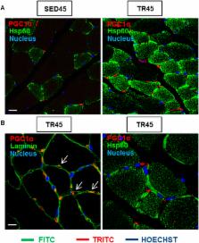
          Heat shock protein 60 (Hsp60) is a chaperone localizing in skeletal muscle mitochondria, whose role is poorly understood. In the present study, the levels of Hsp60 in fibres of the entire posterior group of hindlimb muscles ( gastrocnemius, soleus, and plantaris) were evaluated in mice after completing a 6-week endurance training program. The correlation between Hsp60 levels and the expression of four isoforms of peroxisome proliferator-activated receptor gamma coactivator 1 alpha (PGC1α) were investigated only in soleus. Short-term overexpression of hsp60, achieved by in vitro plasmid transfection, was then performed to determine whether this chaperone could have a role in the activation of the expression levels of PGC1α isoforms. The levels of Hsp60 protein were fibre-type specific in the posterior muscles and endurance training increased its content in type I muscle fibers. Concomitantly with the increased levels of Hsp60 released in the blood stream of trained mice, mitochondrial copy number and the expression of three isoforms of PGC1α increased. Overexpressing hsp60 in cultured myoblasts induced only the expression of PGC1 1α, suggesting a correlation between Hsp60 overexpression and PGC1 1 α activation.

          Related collections

          Most cited references50

          • Record: found
          • Abstract: found
          • Article: not found

          High Levels of Exosomes Expressing CD63 and Caveolin-1 in Plasma of Melanoma Patients

          Background Metastatic melanoma is an untreatable cancer lacking reliable and non-invasive markers of disease progression. Exosomes are small vesicles secreted by normal as well as tumor cells. Human tumor-derived exosomes are involved in malignant progression and we evaluated the presence of exosomes in plasma of melanoma patients as a potential tool for cancer screening and follow-up. Methodology/Principal Findings We designed an in-house sandwich ELISA (Exotest) to capture and quantify exosomes in plasma based on expression of housekeeping proteins (CD63 and Rab-5b) and a tumor-associated marker (caveolin-1). Western blot and flow cytometry analysis of exosomes were used to confirm the Exotest-based findings. The Exotest allowed sensitive detection and quantification of exosomes purified from human tumor cell culture supernatants and plasma from SCID mice engrafted with human melanoma. Plasma levels of exosomes in melanoma-engrafted SCID mice correlated to tumor size. We evaluated the levels of plasma exosomes expressing CD63 and caveolin-1 in melanoma patients (n = 90) and healthy donors (n = 58). Consistently, plasma exosomes expressing CD63 (504±315) or caveolin-1 (619±310) were significantly increased in melanoma patients as compared to healthy donors (223±125 and 228±102, respectively). While the Exotest for CD63+ plasma exosomes had limited sensitivity (43%) the Exotest for detection of caveolin-1+ plasma exosomes showed a higher sensitivity (68%). Moreover, caveolin-1+ plasma exosomes were significantly increased with respect to CD63+ exosomes in the patients group. Conclusions/Significance We describe a new non-invasive assay allowing detection and quantification of human exosomes in plasma of melanoma patients. Our results suggest that the Exotest for detection of plasma exosomes carrying tumor-associated antigens may represent a novel tool for clinical management of cancer patients.
            Bookmark
            • Record: found
            • Abstract: found
            • Article: not found

            A PGC-1α isoform induced by resistance training regulates skeletal muscle hypertrophy.

            PGC-1α is a transcriptional coactivator induced by exercise that gives muscle many of the best known adaptations to endurance-type exercise but has no effects on muscle strength or hypertrophy. We have identified a form of PGC-1α (PGC-1α4) that results from alternative promoter usage and splicing of the primary transcript. PGC-1α4 is highly expressed in exercised muscle but does not regulate most known PGC-1α targets such as the mitochondrial OXPHOS genes. Rather, it specifically induces IGF1 and represses myostatin, and expression of PGC-1α4 in vitro and in vivo induces robust skeletal muscle hypertrophy. Importantly, mice with skeletal muscle-specific transgenic expression of PGC-1α4 show increased muscle mass and strength and dramatic resistance to the muscle wasting of cancer cachexia. Expression of PGC-1α4 is preferentially induced in mouse and human muscle during resistance exercise. These studies identify a PGC-1α protein that regulates and coordinates factors involved in skeletal muscle hypertrophy. Copyright © 2012 Elsevier Inc. All rights reserved.
              Bookmark
              • Record: found
              • Abstract: found
              • Article: not found

              Exercise-induced mitochondrial biogenesis begins before the increase in muscle PGC-1alpha expression.

              Exercise results in rapid increases in expression of the transcription coactivator peroxisome proliferator-activated receptor gamma coactivator-1alpha (PGC-1alpha) and in mitochondrial biogenesis in skeletal muscle. PGC-1alpha regulates and coordinates mitochondrial biogenesis, and overexpression of PGC-1alpha in muscle cells results in increases in mitochondrial content. In this context, it has been proposed that the increase in PGC-1alpha protein expression mediates the exercise-induced increase in mitochondrial biogenesis. However, we found that mitochondrial proteins with a short half-life increase as rapidly as, or more rapidly than, PGC-1alpha protein. This finding led us to hypothesize that activation, rather than increased expression, of PGC-1alpha mediates the initial phase of the exercise-induced increase in mitochondria. In this study, we found that most of the PGC-1alpha in resting skeletal muscle is in the cytosol. Exercise resulted in activation of p38 MAPK and movement of PGC-1alpha into the nucleus. In support of our hypothesis, binding of the transcription factor nuclear respiratory factor 1 (NRF-1) to the cytochrome c promoter and NRF-2 to the cytochrome oxidase subunit 4 promoter increased in response to exercise prior to an increase in PGC-1alpha protein. Furthermore, exercise-induced increases in the mRNAs of cytochrome c, delta-aminolevulinate synthase, and citrate synthase also occurred before an increase in PGC-1 protein. Thus, it appears that activation of PGC-1alpha may mediate the initial phase of the exercise-induced adaptive increase in muscle mitochondria, whereas the subsequent increase in PGC-1alpha protein sustains and enhances the increase in mitochondrial biogenesis.
                Bookmark

                Author and article information

                Journal
                Sci Rep
                Sci Rep
                Scientific Reports
                Nature Publishing Group
                2045-2322
                27 January 2016
                2016
                : 6
                : 19781
                Affiliations
                [1 ]Department of Experimental Biomedicine and Clinical Neurosciences (BioNeC), University of Palermo , Palermo, 90127 Italy
                [2 ]Euro-Mediterranean Institute of Science and Technology (IEMEST) , Palermo, 90100, Italy
                [3 ]Department of Anatomical, Histological, Forensic & Orthopaedic Sciences, Section of Histology & Medical Embryology, Sapienza University of Rome , Rome, 00161, Italy
                [4 ]Interuniversity Institute of Myology , Rome, 00161, Italy
                [5 ]University Pierre et Marie Curie Paris, UR4 Aging, Stress, Inflammation , 75005 Paris, France
                [6 ]Department of Microbiology and Immunology, School of Medicine, University of Maryland at Baltimore, IMET , 21201 Baltimore, MD, USA
                Author notes
                Article
                srep19781
                10.1038/srep19781
                4728392
                26812922
                e6d2f73d-6748-4d7e-a2c4-4d1120de6923
                Copyright © 2016, Macmillan Publishers Limited

                This work is licensed under a Creative Commons Attribution 4.0 International License. The images or other third party material in this article are included in the article’s Creative Commons license, unless indicated otherwise in the credit line; if the material is not included under the Creative Commons license, users will need to obtain permission from the license holder to reproduce the material. To view a copy of this license, visit http://creativecommons.org/licenses/by/4.0/

                History
                : 16 June 2015
                : 17 December 2015
                Categories
                Article

                Uncategorized
                Uncategorized

                Comments

                Comment on this article