8
views
0
recommends
+1 Recommend
0 collections
    0
    shares
      • Record: found
      • Abstract: found
      • Article: found
      Is Open Access

      Prognostic Value of Bone Mineral Density on Curve Progression: A Longitudinal Cohort Study of 513 Girls with Adolescent Idiopathic Scoliosis

      research-article

      Read this article at

      Bookmark
          There is no author summary for this article yet. Authors can add summaries to their articles on ScienceOpen to make them more accessible to a non-specialist audience.

          Abstract

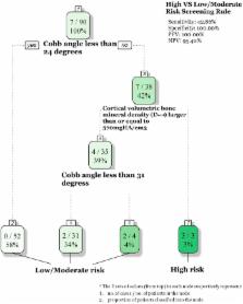
          Osteopenia has been found to occur in about 30% of Adolescent Idiopathic Scoliosis (AIS) patients. This study aimed to investigate its prognostic value on the risk of curve progression to surgical threshold. Newly diagnosed AIS girls (N = 513) with Cobb angle 10°–40° were recruited with follow-up till maturity. Bilateral hips were assessed with dual-energy x-ray absorptiometry (DXA). Distal radius of a subgroup of 90 subjects was further assessed with high-resolution peripheral quantitative computed tomography (HR-pQCT). 55 patients progressed to surgical threshold or underwent spine surgery at the end of follow-up. Cox model with osteopenia status performed significantly better than the model without (p = 0.010). Osteopenic patients had significantly higher risk of surgery (HR2.25, p = 0.011), even after adjustment for menarche status, age and initial Cobb angle. The incremental predictive value of osteopenia was, however, not statistically significant. In the subgroup analysis, cortical bone density was identified as a better marker to improve the sensitivity of the prediction, but requires further larger study to validate this finding. These consistent results of bone density measured at different sites suggest a systemic effect, rather than local effect to the deformed spine, and support to the link of abnormal bone density to the etiopathogenesis in AIS patients.

          Related collections

          Most cited references23

          • Record: found
          • Abstract: found
          • Article: not found

          Adolescent idiopathic scoliosis.

          Adolescent idiopathic scoliosis (AIS) affects 1-3% of children in the at-risk population of those aged 10-16 years. The aetiopathogensis of this disorder remains unknown, with misinformation about its natural history. Non-surgical treatments are aimed to reduce the number of operations by preventing curve progression. Although bracing and physiotherapy are common treatments in much of the world, their effectiveness has never been rigorously assessed. Technological advances have much improved the ability of surgeons to safely correct the deformity while maintaining sagittal and coronal balance. However, we do not have long-term results of these changing surgical treatments. Much has yet to be learned about the general health, quality of life, and self-image of both treated and untreated patients with AIS.
            Bookmark
            • Record: found
            • Abstract: found
            • Article: not found

            Effects of bracing in adolescents with idiopathic scoliosis.

            The role of bracing in patients with adolescent idiopathic scoliosis who are at risk for curve progression and eventual surgery is controversial. We conducted a multicenter study that included patients with typical indications for bracing due to their age, skeletal immaturity, and degree of scoliosis. Both a randomized cohort and a preference cohort were enrolled. Of 242 patients included in the analysis, 116 were randomly assigned to bracing or observation, and 126 chose between bracing and observation. Patients in the bracing group were instructed to wear the brace at least 18 hours per day. The primary outcomes were curve progression to 50 degrees or more (treatment failure) and skeletal maturity without this degree of curve progression (treatment success). The trial was stopped early owing to the efficacy of bracing. In an analysis that included both the randomized and preference cohorts, the rate of treatment success was 72% after bracing, as compared with 48% after observation (propensity-score-adjusted odds ratio for treatment success, 1.93; 95% confidence interval [CI], 1.08 to 3.46). In the intention-to-treat analysis, the rate of treatment success was 75% among patients randomly assigned to bracing, as compared with 42% among those randomly assigned to observation (odds ratio, 4.11; 95% CI, 1.85 to 9.16). There was a significant positive association between hours of brace wear and rate of treatment success (P<0.001). Bracing significantly decreased the progression of high-risk curves to the threshold for surgery in patients with adolescent idiopathic scoliosis. The benefit increased with longer hours of brace wear. (Funded by the National Institute of Arthritis and Musculoskeletal and Skin Diseases and others; BRAIST ClinicalTrials.gov number, NCT00448448.).
              Bookmark
              • Record: found
              • Abstract: found
              • Article: not found

              Adolescent idiopathic scoliosis.

              Adolescent idiopathic scoliosis (AIS) is the most common form of structural spinal deformities that have a radiological lateral Cobb angle - a measure of spinal curvature - of ≥10(°). AIS affects between 1% and 4% of adolescents in the early stages of puberty and is more common in young women than in young men. The condition occurs in otherwise healthy individuals and currently has no recognizable cause. In the past few decades, considerable progress has been made towards understanding the clinical patterns and the three-dimensional pathoanatomy of AIS. Advances in biomechanics and technology and their clinical application, supported by limited evidence-based research, have led to improvements in the safety and outcomes of surgical and non-surgical treatments. However, the definite aetiology and aetiopathogenetic mechanisms that underlie AIS are still unclear. Thus, at present, both the prevention of AIS and the treatment of its direct underlying cause are not possible.
                Bookmark

                Author and article information

                Journal
                Sci Rep
                Sci Rep
                Scientific Reports
                Nature Publishing Group
                2045-2322
                19 December 2016
                2016
                : 6
                : 39220
                Affiliations
                [1 ]Department of Orthopaedics and Traumatology, Faculty of Medicine, Prince of Wales Hospital, The Chinese University of Hong Kong , Hong Kong
                [2 ]JC School of Public Health and Primary Care, Faculty of Medicine, The Chinese University of Hong Kong , Hong Kong
                [3 ]Joint Scoliosis Research Center of the Chinese University of Hong Kong and Nanjing University , Hong Kong
                [4 ]Bone Quality and Health Centre, Department of Orthopaedics and Traumatology, Faculty of Medicine, Prince of Wales Hospital, The Chinese University of Hong Kong , Hong Kong
                [5 ]SH Ho Scoliosis Research Laboratory, Faculty of Medicine, Prince of Wales Hospital, The Chinese University of Hong Kong , Hong Kong
                [6 ]Department of Orthopedic Surgery, Second Affiliated Hospital, School of Medicine, Zhejiang University, Hangzhou , China
                [7 ]Spine Surgery, the Affiliated Drum Tower Hospital of Nanjing University Medical School , Nanjing, China
                Author notes
                [*]

                These authors contributed equally to this work.

                Article
                srep39220
                10.1038/srep39220
                5171643
                27991528
                e5d2c325-95ed-4e49-9773-3f5cc63f7659
                Copyright © 2016, The Author(s)

                This work is licensed under a Creative Commons Attribution 4.0 International License. The images or other third party material in this article are included in the article’s Creative Commons license, unless indicated otherwise in the credit line; if the material is not included under the Creative Commons license, users will need to obtain permission from the license holder to reproduce the material. To view a copy of this license, visit http://creativecommons.org/licenses/by/4.0/

                History
                : 17 August 2016
                : 21 November 2016
                Categories
                Article

                Uncategorized
                Uncategorized

                Comments

                Comment on this article