8
views
0
recommends
+1 Recommend
0 collections
    0
    shares
      • Record: found
      • Abstract: found
      • Article: found
      Is Open Access

      Therapeutic Potential of Sclareol in Experimental Models of Rheumatoid Arthritis

      research-article

      Read this article at

      Bookmark
          There is no author summary for this article yet. Authors can add summaries to their articles on ScienceOpen to make them more accessible to a non-specialist audience.

          Abstract

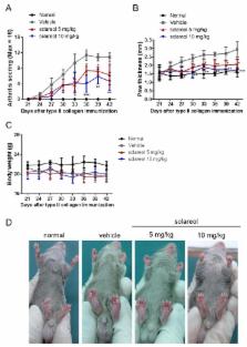
          Previous studies have shown that the natural diterpene compound, sclareol, potentially inhibits inflammation, but it has not yet been determined whether sclareol can alleviate inflammation associated with rheumatoid arthritis (RA). Here, we utilized human synovial cell line, SW982, and an experimental murine model of rheumatoid arthritis, collagen-induced arthritis (CIA), to evaluate the therapeutic effects of sclareol in RA. Arthritic DBA/1J mice were dosed with 5 and 10 mg/kg sclareol intraperitoneally every other day over 21 days. Arthritic severity was evaluated by levels of anti-collagen II (anti-CII) antibody, inflammatory cytokines, and histopathologic examination of knee joint tissues. Our results reveal that the serum anti-CII antibody, cytokines interleukin (IL)-1β, IL-6, tumor necrosis factor (TNF)-α, and IL-17, as well as Th17 and Th1 cell population in inguinal lymph nodes, were significantly lower in sclareol-treated mice compared to the control group. Also, the sclareol treatment groups showed reduced swelling in the paws and lower histological arthritic scores, indicating that sclareol potentially mitigates collagen-induced arthritis. Furthermore, IL-1β-stimulated SW982 cells secreted less inflammatory cytokines (TNF-α and IL-6), which is associated with the downregulation of p38-mitogen-activated protein kinase (MAPK), extracellular signal-regulated kinase (ERK), and NF-κB pathways. Overall, we demonstrate that sclareol could relieve arthritic severities by modulating excessive inflammation and our study merits the pharmaceutical development of sclareol as a therapeutic treatment for inflammation associated with RA.

          Related collections

          Most cited references28

          • Record: found
          • Abstract: found
          • Article: not found

          Fibroblast-like synoviocytes: key effector cells in rheumatoid arthritis.

          Rheumatoid arthritis (RA) remains a significant unmet medical need despite significant therapeutic advances. The pathogenesis of RA is complex and includes many cell types, including T cells, B cells, and macrophages. Fibroblast-like synoviocytes (FLS) in the synovial intimal lining also play a key role by producing cytokines that perpetuate inflammation and proteases that contribute to cartilage destruction. Rheumatoid FLS develop a unique aggressive phenotype that increases invasiveness into the extracellular matrix and further exacerbates joint damage. Recent advances in understanding the biology of FLS, including their regulation regulate innate immune responses and activation of intracellular signaling mechanisms that control their behavior, provide novel insights into disease mechanisms. New agents that target FLS could potentially complement the current therapies without major deleterious effect on adaptive immune responses.
            Bookmark
            • Record: found
            • Abstract: found
            • Article: found
            Is Open Access

            Fibroblast-like synoviocyte metabolism in the pathogenesis of rheumatoid arthritis

            An increasing number of studies show how changes in intracellular metabolic pathways alter tumor and immune cell function. However, little information about metabolic changes in other cell types, including synovial fibroblasts, is available. In rheumatoid arthritis (RA), fibroblast-like synoviocytes (FLS) are the most common cell type at the pannus–cartilage junction and contribute to joint destruction through their production of cytokines, chemokines, and matrix-degrading molecules and by migrating and invading joint cartilage. In this review, we show that these cells differ from healthy synovial fibroblasts, not only in their marker expression, proto-oncogene expression, or their epigenetic changes, but also in their intracellular metabolism. These metabolic changes must occur due to the stressful microenvironment of inflamed tissues, where concentrations of crucial nutrients such as glucose, glutamine, and oxygen are spatially and temporally heterogeneous. In addition, these metabolic changes will increase metabolite exchange between fibroblast and other synovial cells, which can potentially be activated. Glucose and phospholipid metabolism as well as bioactive lipids, including sphingosine-1-phosphate and lysophosphatidic acid, among others, are involved in FLS activation. These metabolic changes likely contribute to FLS involvement in aspects of immune response initiation or abnormal immune responses and strongly contribute to joint destruction.
              Bookmark
              • Record: found
              • Abstract: found
              • Article: found
              Is Open Access

              IL-17-mediated mitochondrial dysfunction impairs apoptosis in rheumatoid arthritis synovial fibroblasts through activation of autophagy

              Fibroblast-like synoviocytes (FLSs) are a major cell population of the pannus that invades cartilage and bone in rheumatoid arthritis (RA). FLS resistance to apoptosis is a major characteristic of RA. The aims of this study were to investigate the effects of interleukin-17 (IL-17) and IL-17-producing T helper (Th17) cells on resistance to apoptosis in FLSs from RA patients (RA FLSs) and their roles in mitochondrial dysfunction and autophagy. Mitochondrial function was assessed in RA FLSs and FLSs from osteoarthritis patients (OA FLSs). FLSs were treated with IL-17 and their morphological features, respiratory level and mitochondrial gene expression were measured. The effects of IL-17 and Th17 cells on the relationship between autophagy and apoptosis were evaluated by measuring the expression of apoptosis-related genes using sodium nitroprusside or 3-methyladenine. The mitochondria of FLSs isolated from RA and osteoarthritis patients displayed different morphological and physiological features. RA FLSs exhibited greater autophagosome formation and greater dysfunction of mitochondrial respiration compared with OA FLSs. IL-17 induced mitochondrial dysfunction and autophagosome formation in RA FLSs, suggesting that they were resistant to apoptosis. Autophagy-related antiapoptosis induced by IL-17 was restored by inhibition of autophagy, suggesting a relationship between mitochondrial dysfunction and cell survival in RA FLSs. Th17 cells and IL-17 increased autophagy of RA FLSs by causing mitochondrial dysfunction. Our findings suggest that, in RA, interactions between RA FLSs and Th17 cells may be involved in the tumorous growth of FLSs and the formation of pannus in joints.
                Bookmark

                Author and article information

                Journal
                Int J Mol Sci
                Int J Mol Sci
                ijms
                International Journal of Molecular Sciences
                MDPI
                1422-0067
                03 May 2018
                May 2018
                : 19
                : 5
                : 1351
                Affiliations
                [1 ]Department of Physical Medicine and Rehabilitation, Taichung Tzu Chi Hospital, Buddhist Tzu Chi Medical Foundation, Taichung 427, Taiwan; tsaisenwei@ 123456gmail.com
                [2 ]Department of Physical Medicine and Rehabilitation, School of Medicine, Tzu Chi University, Hualien 970, Taiwan
                [3 ]Division of Endocrinology and Metabolism, Department of Internal Medicine, Changhua Christian Hospital, Changhua 500, Taiwan; mingchia570531@ 123456gmail.com
                [4 ]Hubei Key Laboratory of Processing and Application of Catalytic Materials, College of Chemical Engineering, Huanggang Normal University, Huanggang 438000, China; shiming3702@ 123456gmail.com
                [5 ]National Center for Biodefense and Infectious Diseases, School of Systems Biology, George Mason University, Manassas, VA 20110, USA; slin20@ 123456gmu.edu (S.-C.L.); cwoodso2@ 123456gmu.edu (C.W.L.)
                [6 ]Institute of Biomedical Science, National Chung-Hsing University, Taichung 40227, Taiwan
                [7 ]Department of Orthopaedics, Taichung Veterans General Hospital, Taichung 40705, Taiwan; wsp0120@ 123456yahoo.com.tw
                [8 ]Biodefense Program, Schar School of Policy and Government, George Mason University, Fairfax, VA 20110, USA; christopher.zane.lien@ 123456gmail.com
                [9 ]Department of Medical Research, China Medical University Hospital, Taichung 40402, Taiwan
                Author notes
                [†]

                These authors contributed equally to this work.

                Author information
                https://orcid.org/0000-0003-2942-5937
                Article
                ijms-19-01351
                10.3390/ijms19051351
                5983692
                29751535
                e51950b7-cc2c-44a9-9e6a-391d7df7ecc1
                © 2018 by the authors.

                Licensee MDPI, Basel, Switzerland. This article is an open access article distributed under the terms and conditions of the Creative Commons Attribution (CC BY) license ( http://creativecommons.org/licenses/by/4.0/).

                History
                : 10 April 2018
                : 26 April 2018
                Categories
                Article

                Molecular biology
                sclareol,rheumatoid arthritis,synovial cell,collagen,mice,cytokines,th17,mapk
                Molecular biology
                sclareol, rheumatoid arthritis, synovial cell, collagen, mice, cytokines, th17, mapk

                Comments

                Comment on this article