31
views
0
recommends
+1 Recommend
0 collections
    0
    shares
      • Record: found
      • Abstract: found
      • Article: found
      Is Open Access

      Enhancement of Force Generated by Individual Myosin Heads in Skinned Rabbit Psoas Muscle Fibers at Low Ionic Strength

      research-article

      Read this article at

      Bookmark
          There is no author summary for this article yet. Authors can add summaries to their articles on ScienceOpen to make them more accessible to a non-specialist audience.

          Abstract

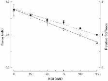
          Although evidence has been presented that, at low ionic strength, myosin heads in relaxed skeletal muscle fibers form linkages with actin filaments, the effect of low ionic strength on contraction characteristics of Ca 2+-activated muscle fibers has not yet been studied in detail. To give information about the mechanism of muscle contraction, we have examined the effect of low ionic strength on the mechanical properties and the contraction characteristics of skinned rabbit psoas muscle fibers in both relaxed and maximally Ca 2+-activated states. By progressively decreasing KCl concentration from 125 mM to 0 mM (corresponding to a decrease in ionic strength μ from 170 mM to 50 mM), relaxed fibers showed changes in mechanical response to sinusoidal length changes and ramp stretches, which are consistent with the idea of actin-myosin linkage formation at low ionic strength. In maximally Ca 2+-activated fibers, on the other hand, the maximum isometric force increased about twofold by reducing KCl concentration from 125 to 0 mM. Unexpectedly, determination of the force-velocity curves indicated that, the maximum unloaded shortening velocity V max, remained unchanged at low ionic strength. This finding indicates that the actin-myosin linkages, which has been detected in relaxed fibers at low ionic strength, are broken quickly on Ca 2+ activation, so that the linkages in relaxed fibers no longer provide any internal resistance against fiber shortening. The force-velocity curves, obtained at various levels of steady Ca 2+-activated isometric force, were found to be identical if they are normalized with respect to the maximum isometric force. The MgATPase activity of muscle fibers during isometric force generation was found not to change appreciably at low ionic strength despite the two-fold increase in Ca 2+-activated isometric force. These results can be explained in terms of enhancement of force generated by individual myosin heads, but not by any changes in kinetic properties of cyclic actin-myosin interaction.

          Related collections

          Most cited references19

          • Record: found
          • Abstract: not found
          • Article: not found

          Mechanism of adenosine triphosphate hydrolysis by actomyosin.

            Bookmark
            • Record: found
            • Abstract: found
            • Article: not found

            X-ray diffraction measurements of the extensibility of actin and myosin filaments in contracting muscle.

            We have used a small angle scattering system assembled on the high flux multipole wiggler beam line at CHESS (Cornell) to make very accurate spacing measurements of certain meridional and layer-line reflections from contracting muscles. During isometric contraction, the actin 27.3 A reflection increases in spacing from its resting value by approximately 0.3%, and other actin reflections, including the 59 and 51 A off-meridional reflections, show corresponding changes in spacing. When tension is augmented or diminished by applying moderate speed length changes to a contracting muscle, changes in spacing in the range of 0.19-0.24% (when scaled to full isometric tension) can be seen. The larger difference between the resting and isometric spacings suggests either nonlinearity at low tension levels or the presence of a component related to activation itself. Myosin filaments also show similar increases in axial period during slow stretch, in addition to the well known larger change associated with activation. An actin spacing change of 0.25-0.3% can also be measured during a 2 ms time frame immediately after a quick release, showing that the elastic behavior is rapid. These observations of filament extensions totaling 2-3 nm per half-sarcomere may necessitate some significant revision of the interpretation of a number of mechanical experiments in muscle, in which it has usually been assumed that virtually all of the elasticity resides in the cross-bridges.
              Bookmark
              • Record: found
              • Abstract: found
              • Article: not found

              Double-hyperbolic force-velocity relation in frog muscle fibres.

              K. Edman (1988)
              1. The relationship between force and velocity of shortening was studied at 2.10 micron sarcomere length during fused tetani (1-3 degrees C) in single fibres isolated from the anterior tibialis muscle of Rana temporaria. The speed of shortening was recorded from the whole fibre and, in some experiments, simultaneously from a short (ca. 0.6 mm) segment, while the preparation was released to shorten isotonically at selected force levels ('load-clamp' recording). The segment was defined by opaque markers of hair that were placed on the fibre surface. The distance between the markers was recorded by means of a photo-electric detector system. 2. The force-velocity relation had two distinct regions, each one exhibiting an upwards concave shape, that were located within the ranges 0-78 and 78-100% of the measured isometric force (P0), respectively. The two portions of the force-velocity relation could be fitted well by hyperbolic functions or by single-exponential functions. The curvature was more pronounced in the high-force region than at low-intermediate loads. The transition between the two portions of the force-velocity relation (the 'break point' of the force-velocity curve) occurred at 78.4 +/- 0.4% of P0 (mean +/- S.E. of mean, n = 12) corresponding to 10.9 +/- 0.4% of maximum velocity of shortening (Vmax). The general shape of the force-velocity curve, and the appearance of a break point near 78% of P0, was the same when measurements were made from the whole fibre and from a short segment along the same fibre. 3. The 'negative' branch of the force-velocity relation was delineated for loads ranging from P0 to 1.6-1.8 P0 in five experiments. The negative branch formed a smooth continuation of the force-velocity relation recorded between 0.78 P0 and P0. The force-velocity relation was nearly flat between 0.90 P0 and 1.20 P0, the difference in speed of shortening or elongation being 1.8 +/- 0.3% (mean +/- S.E. of mean, n = 5) of Vmax over this range. 4. An increase in sarcomere length from 1.85 to 2.60 micron did not affect Vmax but caused a steady decrease in curvature of the force-velocity relation, both at low-intermediate loads and in the high-force range. Similar changes in shape of the force-velocity relation were produced by osmotic compression of the fibre in a Ringer solution made hypertonic by addition of 98 mM-sucrose.(ABSTRACT TRUNCATED AT 400 WORDS)
                Bookmark

                Author and article information

                Contributors
                Role: Editor
                Journal
                PLoS One
                PLoS ONE
                plos
                plosone
                PLoS ONE
                Public Library of Science (San Francisco, USA )
                1932-6203
                2013
                15 May 2013
                : 8
                : 5
                : e63658
                Affiliations
                [1 ]Department of Physiology, School of Medicine, Teikyo University, Tokyo, Japan
                [2 ]Department of Electronic Engineering, Shibaura Institute of Technology, Tokyo, Japan
                [3 ]Department of Integrated Sciences in Physics and Biology, College of Humanities and Sciences, Nihon University, Tokyo, Japan
                [4 ]Department of Physiology, School of Dentistry, Tsurumi University, Yokohama, Japan
                [5 ]Graduate School of Frontier Sciences, University of Tokyo, Tokyo, Japan
                Cinvestav-IPN, Mexico
                Author notes

                Competing Interests: The authors have declared that no competing interests exist.

                Conceived and designed the experiments: HS TK SC. Performed the experiments: HS TA YO YS SS. Analyzed the data: HS TA TK SC YS. Wrote the paper: HS TK SC SS. Created the figures: TA YO.

                Article
                PONE-D-13-01089
                10.1371/journal.pone.0063658
                3655179
                23691080
                e4adaaf1-3c7c-4190-97e5-d763c68de6ba
                Copyright @ 2013

                This is an open-access article distributed under the terms of the Creative Commons Attribution License, which permits unrestricted use, distribution, and reproduction in any medium, provided the original author and source are credited.

                History
                : 30 December 2012
                : 4 April 2013
                Page count
                Pages: 8
                Funding
                The authors have no support or funding to report.
                Categories
                Research Article
                Biology
                Anatomy and Physiology
                Musculoskeletal System
                Muscle
                Muscle Biochemistry
                Biochemistry
                Proteins
                Contractile Proteins
                Molecular Cell Biology
                Cellular Types
                Muscle Fibers
                Medicine
                Anatomy and Physiology
                Musculoskeletal System
                Muscle
                Muscle Biochemistry

                Uncategorized
                Uncategorized

                Comments

                Comment on this article