5
views
0
recommends
+1 Recommend
0 collections
    0
    shares
      • Record: found
      • Abstract: found
      • Article: found
      Is Open Access

      Mechanical overloading induces GPX4-regulated chondrocyte ferroptosis in osteoarthritis via Piezo1 channel facilitated calcium influx

      research-article
      a , b , c , d , 1 , e , 1 , a , f , b , f , a , g , h , i , j , k , l , l , m , n , a , a , a , b , * , a , *
      Journal of Advanced Research
      Elsevier
      Chondrocytes, Osteoarthritis, Ferroptosis, Mechanical stress, Piezo1, GPX4, Glutathione peroxidase 4, OA, osteoarthritis, CoQ10, coenzyme Q10, FSP-1, ferroptosis suppressor protein 1, GSH, glutathione, DMM, Destabilization of the medial meniscus, MMP-13, Matrix metallopeptidase 13, ADAMTS-5, A disintegrin and metalloproteinase with thrombospondin motifs-5, Col2, Collagen II, ROS, Reactive oxygen species, GAG, Glycosaminoglycan, OARSI, Osteoarthritis Research Society International

      Read this article at

      Bookmark
          There is no author summary for this article yet. Authors can add summaries to their articles on ScienceOpen to make them more accessible to a non-specialist audience.

          Graphical abstract

          Highlight:

          • Our study proved that mechanical overloading induces ferroptosis of chondrocyte, which might be a potential therapeutic target for mechanical damage of chondrocyte and OA.

          • Our study demonstrated Piezo1 facilitated calcium influx leads to reduction of GSH, decrease of Gpx4 and activation of oxidative stress in chondrocyte under high strain mechanical stimulation.

          • Mechanical signals were converted into ferroptosis-associated signals through Piezo1 channel induced calcium influx, which might shed light on therapeutic interventions for treatment of OA and other diseases associated with ferroptosis.

          Abstract

          Introductions

          Excessive mechanical stress is closely associated with cell death in various conditions. Exposure of chondrocytes to excessive mechanical loading leads to a catabolic response as well as exaggerated cell death. Ferroptosis is a recently identified form of cell death during cell aging and degeneration. However, it's potential association with mechanical stress remains to be illustrated.

          Objectives

          To identify whether excessive mechanical stress can cause ferroptosis. To explore the role of mechanical overloading in chondrocyte ferroptosis.

          Methods

          Chondrocytes were collected from loading and unloading zones of cartilage in patients with osteoarthritis (OA), and the ferroptosis phenotype was analyzed through transmission electron microscope and microarray. Moreover, the relationship between ferroptosis and OA was analyzed by GPX4-conditional knockout (Col2a1-CreERT: GPX4 flox/flox) mice OA model and chondrocytes cultured with high strain mechanical stress. Furthermore, the role of Piezo1 ion channel in chondrocyte ferroptosis and OA development was explored by using its inhibitor (GsMTx4) and agonist (Yoda1). Additionally, chondrocyte was cultured in calcium-free medium with mechanical stress, and ferroptosis phenotype was tested.

          Results

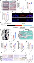
          Human cartilage and mouse chondrocyte experiments revealed that mechanical overloading can induce GPX4-associated ferroptosis. Conditional knockout of GPX4 in cartilage aggravated experimental OA process, while additional treatment with ferroptosis suppressor protein (FSP-1) and coenzyme Q10 (CoQ10) abated OA development in GPX4-CKO mice. In mouse OA model and chondrocyte experiments, inhibition of Piezo1 channel activity increased GPX4 expression, attenuated ferroptosis phenotype and reduced the severity of osteoarthritis. Additionally, high strain mechanical stress induced ferroptosis damage in chondrocyte was largely abolished by blocking calcium influx through calcium-free medium.

          Conclusions

          Our findings show that mechanical overloading induces ferroptosis through Piezo1 activation and subsequent calcium influx in chondrocytes, which might provide a potential target for OA treatment .

          Related collections

          Most cited references52

          • Record: found
          • Abstract: found
          • Article: not found

          The CoQ oxidoreductase FSP1 acts in parallel to GPX4 to inhibit ferroptosis

          Ferroptosis is a form of regulated cell death that is caused by the iron-dependent peroxidation of lipids 1,2 . The glutathione-dependent lipid hydroperoxidase glutathione peroxidase 4 (GPX4) prevents ferroptosis by converting lipid hydroperoxides into non-toxic lipid alcohols 3,4 . Ferroptosis has been implicated in the cell death that underlies several degenerative conditions 2 , and induction of ferroptosis by inhibition of GPX4 has emerged as a therapeutic strategy to trigger cancer cell death 5 . However, sensitivity to GPX4 inhibitors varies greatly across cancer cell lines 6 , suggesting that additional factors govern resistance to ferroptosis. Here, employing a synthetic lethal CRISPR/Cas9 screen, we identify ferroptosis suppressor protein 1 (FSP1) (previously known as apoptosis-inducing factor mitochondrial 2 (AIFM2)) as a potent ferroptosis resistance factor. Our data indicate that myristoylation recruits FSP1 to the plasma membrane where it functions as an oxidoreductase that reduces coenzyme Q10 (CoQ), generating a lipophilic radical-trapping antioxidant (RTA) that halts the propagation of lipid peroxides. We further find that FSP1 expression positively correlates with ferroptosis resistance across hundreds of cancer cell lines, and that FSP1 mediates resistance to ferroptosis in lung cancer cells in culture and in mouse tumor xenografts. Thus, our data identify FSP1 as a key component of a non-mitochondrial CoQ antioxidant system that acts in parallel to the canonical glutathione-based GPX4 pathway. These findings define a new ferroptosis suppression pathway and indicate that pharmacological inhibition of FSP1 may provide an effective strategy to sensitize cancer cells to ferroptosis-inducing chemotherapeutics.
            Bookmark
            • Record: found
            • Abstract: not found
            • Article: not found

            FSP1 is a glutathione-independent ferroptosis suppressor

              Bookmark
              • Record: found
              • Abstract: found
              • Article: not found

              Broadening horizons: the role of ferroptosis in cancer

              The discovery of regulated cell death processes has enabled advances in cancer treatment. In the past decade, ferroptosis, an iron-dependent form of regulated cell death driven by excessive lipid peroxidation, has been implicated in the development and therapeutic responses of various types of tumours. Experimental reagents (such as erastin and RSL3), approved drugs (for example, sorafenib, sulfasalazine, statins and artemisinin), ionizing radiation and cytokines (such as IFNγ and TGFβ1) can induce ferroptosis and suppress tumour growth. However, ferroptotic damage can trigger inflammation-associated immunosuppression in the tumour microenvironment, thus favouring tumour growth. The extent to which ferroptosis affects tumour biology is unclear, although several studies have found important correlations between mutations in cancer-relevant genes (for example, RAS and TP53), in genes encoding proteins involved in stress response pathways (such as NFE2L2 signalling, autophagy and hypoxia) and the epithelial-to-mesenchymal transition, and responses to treatments that activate ferroptosis. Herein, we present the key molecular mechanisms of ferroptosis, describe the crosstalk between ferroptosis and tumour-associated signalling pathways, and discuss the potential applications of ferroptosis in the context of systemic therapy, radiotherapy and immunotherapy.
                Bookmark

                Author and article information

                Contributors
                Journal
                J Adv Res
                J Adv Res
                Journal of Advanced Research
                Elsevier
                2090-1232
                2090-1224
                11 January 2022
                November 2022
                11 January 2022
                : 41
                : 63-75
                Affiliations
                [a ]Department of Orthopaedic Surgery, Qilu Hospital, Cheeloo College of Medicine, Shandong University, Jinan, Shandong 250012, PR China
                [b ]Qilu Hospital of Shandong University Spine and Spinal Cord Disease Research Center-ICMRS Collaborating Center for Orthopaedic translational Research, Shandong University, Jinan, Shandong 250012, PR China
                [c ]Institute of Stomatology, Shandong University, Jinan, Shandong, 250012, PR China
                [d ]NHC Key Laboratory of Otorhinolaryngology, Qilu Hospital, Cheeloo College of Medicine, Shandong University, Jinan, Shandong 250012, PR China
                [e ]Department of Pathology, Qilu Hospital, Cheeloo College of Medicine, Shandong University, Jinan, Shandong 250012, PR China
                [f ]Cheeloo College of Medicine, Shandong University, Jinan, Shandong, 250012, PR China
                [g ]Department of Orthopaedic Surgery, New York University School of Medicine, New York University Medical Center, New York City, NY, USA
                [h ]Future Industries Institute, University of South Australia, Mawson Lakes Campus, Mawson Lakes 5095, Australia
                [i ]School of Engineering, University of South Australia, Mawson Lakes Campus, Mawson Lakes 5095, Australia
                [j ]Department of Orthopaedic Surgery, The First Affiliated Hospital of Shandong First Medical University, Jinan, Shandong 250012, PR China
                [k ]Health Management Centre. The First Affiliated Hospital of Shandong First Medical University, Jinan, Shandong 250012, PR China
                [l ]Experimental Therapeutics Laboratory, University of South Australia Cancer Research Institute, Adelaide SA 5000, Australia
                [m ]Robinson Research Institute and Adelaide Medical School, University of Adelaide, Adelaide SA 5005, Australia
                [n ]Key Laboratory of Colloid and Interface Chemistry (Shandong University), Ministry of Education, Jinan 250100, PR China
                Author notes
                [* ]Corresponding authors at: Qilu Hospital, Cheeloo College of Medicine, Shandong University, 107 Wenhuaxi Road, Jinan, PR China. chenglei@ 123456email.sdu.edu.cn lwwzyp@ 123456email.sdu.edu.cn
                [1]

                These authors contributed equally to this work.

                Article
                S2090-1232(22)00004-2
                10.1016/j.jare.2022.01.004
                9637484
                36328754
                e3907a00-1602-4a4e-88ad-c9506ed32147
                © 2022 The Authors. Published by Elsevier B.V. on behalf of Cairo University.

                This is an open access article under the CC BY-NC-ND license (http://creativecommons.org/licenses/by-nc-nd/4.0/).

                History
                : 3 October 2021
                : 19 December 2021
                : 7 January 2022
                Categories
                Original Article

                chondrocytes,osteoarthritis,ferroptosis,mechanical stress,piezo1,gpx4, glutathione peroxidase 4,oa, osteoarthritis,coq10, coenzyme q10,fsp-1, ferroptosis suppressor protein 1,gsh, glutathione,dmm, destabilization of the medial meniscus,mmp-13, matrix metallopeptidase 13,adamts-5, a disintegrin and metalloproteinase with thrombospondin motifs-5,col2, collagen ii,ros, reactive oxygen species,gag, glycosaminoglycan,oarsi, osteoarthritis research society international

                Comments

                Comment on this article