3
views
0
recommends
+1 Recommend
0 collections
    0
    shares
      • Record: found
      • Abstract: found
      • Article: found
      Is Open Access

      Membrane curvature induces cardiolipin sorting

      research-article

      Read this article at

      Bookmark
          There is no author summary for this article yet. Authors can add summaries to their articles on ScienceOpen to make them more accessible to a non-specialist audience.

          Abstract

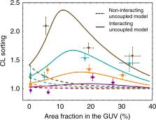
          Cardiolipin is a cone-shaped lipid predominantly localized in curved membrane sites of bacteria and in the mitochondrial cristae. This specific localization has been argued to be geometry-driven, since the CL’s conical shape relaxes curvature frustration. Although previous evidence suggests a coupling between CL concentration and membrane shape in vivo, no precise experimental data are available for curvature-based CL sorting in vitro. Here, we test this hypothesis in experiments that isolate the effects of membrane curvature in lipid-bilayer nanotubes. CL sorting is observed with increasing tube curvature, reaching a maximum at optimal CL concentrations, a fact compatible with self-associative clustering. Observations are compatible with a model of membrane elasticity including van der Waals entropy, from which a negative intrinsic curvature of −1.1 nm −1 is predicted for CL. The results contribute to understanding the physicochemical interplay between membrane curvature and composition, providing key insights into mitochondrial and bacterial membrane organization and dynamics.

          Abstract

          Elena Beltrán-Heredia, Feng-Ching Tsai et al. examine the role of cardiolipin in membrane curvature, finding evidence compatible with a model based on membrane elasticity and van der Waals entropy. These results promote understanding of the interplay between membrane curvature and composition.

          Related collections

          Most cited references28

          • Record: found
          • Abstract: found
          • Article: not found

          Aggregation and vesiculation of membrane proteins by curvature-mediated interactions.

          Membrane remodelling plays an important role in cellular tasks such as endocytosis, vesiculation and protein sorting, and in the biogenesis of organelles such as the endoplasmic reticulum or the Golgi apparatus. It is well established that the remodelling process is aided by specialized proteins that can sense as well as create membrane curvature, and trigger tubulation when added to synthetic liposomes. Because the energy needed for such large-scale changes in membrane geometry significantly exceeds the binding energy between individual proteins and between protein and membrane, cooperative action is essential. It has recently been suggested that curvature-mediated attractive interactions could aid cooperation and complement the effects of specific binding events on membrane remodelling. But it is difficult to experimentally isolate curvature-mediated interactions from direct attractions between proteins. Moreover, approximate theories predict repulsion between isotropically curving proteins. Here we use coarse-grained membrane simulations to show that curvature-inducing model proteins adsorbed on lipid bilayer membranes can experience attractive interactions that arise purely as a result of membrane curvature. We find that once a minimal local bending is realized, the effect robustly drives protein cluster formation and subsequent transformation into vesicles with radii that correlate with the local curvature imprint. Owing to its universal nature, curvature-mediated attraction can operate even between proteins lacking any specific interactions, such as newly synthesized and still immature membrane proteins in the endoplasmic reticulum.
            Bookmark
            • Record: found
            • Abstract: found
            • Article: not found

            Structure of fully hydrated fluid phase lipid bilayers with monounsaturated chains.

            Quantitative structures are obtained at 30 degrees C for the fully hydrated fluid phases of palmitoyloleoylphosphatidylcholine (POPC), with a double bond on the sn-2 hydrocarbon chain, and for dierucoylphosphatidylcholine (di22:1PC), with a double bond on each hydrocarbon chain. The form factors F(qz) for both lipids are obtained using a combination of three methods. (1) Volumetric measurements provide F(0). (2) X-ray scattering from extruded unilamellar vesicles provides /F(qz)/ for low q(z). (3) Diffuse X-ray scattering from oriented stacks of bilayers provides /F(qz)/ for high q(z). Also, data using method (2) are added to our recent data for dioleoylphosphatidylcholine (DOPC) using methods (1) and (3); the new DOPC data agree very well with the recent data and with (4) our older data obtained using a liquid crystallographic X-ray method. We used hybrid electron density models to obtain structural results from these form factors. The result for area per lipid (A) for DOPC 72.4 +/- 0.5 A(2) agrees well with our earlier publications, and we find A = 69.3 +/- 0.5 A2 for di22:1PC and A = 68.3 +/- 1.5 A2 for POPC. We obtain the values for five different average thicknesses: hydrophobic, steric, head-head, phosphate-phosphate and Luzzati. Comparison of the results for these three lipids and for our recent dimyristoylphosphatidylcholine (DMPC) determination provides quantitative measures of the effect of unsaturation on bilayer structure. Our results suggest that lipids with one monounsaturated chain have quantitative bilayer structures closer to lipids with two monounsaturated chains than to lipids with two completely saturated chains.
              Bookmark
              • Record: found
              • Abstract: found
              • Article: not found

              Dynamic subcompartmentalization of the mitochondrial inner membrane

              The inner membrane of mitochondria is organized in two morphologically distinct domains, the inner boundary membrane (IBM) and the cristae membrane (CM), which are connected by narrow, tubular cristae junctions. The protein composition of these domains, their dynamics, and their biogenesis and maintenance are poorly understood at the molecular level. We have used quantitative immunoelectron microscopy to determine the distribution of a collection of representative proteins in yeast mitochondria belonging to seven major processes: oxidative phosphorylation, protein translocation, metabolite exchange, mitochondrial morphology, protein translation, iron–sulfur biogenesis, and protein degradation. We show that proteins are distributed in an uneven, yet not exclusive, manner between IBM and CM. The individual distributions reflect the physiological functions of proteins. Moreover, proteins can redistribute between the domains upon changes of the physiological state of the cell. Impairing assembly of complex III affects the distribution of partially assembled subunits. We propose a model for the generation of this dynamic subcompartmentalization of the mitochondrial inner membrane.
                Bookmark

                Author and article information

                Contributors
                francao@ucm.es
                patricia.bassereau@curie.fr
                monroy@ucm.es
                Journal
                Commun Biol
                Commun Biol
                Communications Biology
                Nature Publishing Group UK (London )
                2399-3642
                20 June 2019
                20 June 2019
                2019
                : 2
                : 225
                Affiliations
                [1 ]ISNI 0000 0001 2157 7667, GRID grid.4795.f, Departamento de Estructura de la Materia, Física Térmica y Electrónica, , Universidad Complutense de Madrid, ; Plaza de Ciencias, 1, 28040 Madrid, Spain
                [2 ]ISNI 0000 0001 2157 7667, GRID grid.4795.f, Departamento de Química Física, , Universidad Complutense de Madrid, ; Avda. Complutense, s/n, 28040 Madrid, Spain
                [3 ]ISNI 0000 0004 1784 3645, GRID grid.440907.e, Laboratoire Physico Chimie Curie, Institut Curie, , PSL Research University, ; CNRS UMR168, 75005 Paris, France
                [4 ]ISNI 0000 0004 0500 5230, GRID grid.429045.e, Instituto Madrileño de Estudios Avanzados en Nanociencia, IMDEA Nanociencia, ; Calle Faraday, 9, 28049 Madrid, Spain
                [5 ]ISNI 0000 0001 2308 1657, GRID grid.462844.8, Sorbonne Université, ; UPMC Univ Paris 06, 75005 Paris, France
                [6 ]ISNI 0000 0001 1945 5329, GRID grid.144756.5, Unit of Translational Biophysics, , Instituto de Investigación Sanitaria Hospital Doce de Octubre (imas12), ; Avda. de Córdoba, s/n, 28041 Madrid, Spain
                [7 ]ISNI 0000 0001 2181 7878, GRID grid.47840.3f, Institute for Quantitative Biosciences-QB3, , University of California at Berkeley, ; Berkeley, CA 94720 USA
                Author information
                http://orcid.org/0000-0003-1611-913X
                http://orcid.org/0000-0002-8544-6778
                http://orcid.org/0000-0001-6455-3083
                Article
                471
                10.1038/s42003-019-0471-x
                6586900
                31240263
                e28efe1d-78cd-4ee2-a02b-0be4bb6e096d
                © The Author(s) 2019

                Open Access This article is licensed under a Creative Commons Attribution 4.0 International License, which permits use, sharing, adaptation, distribution and reproduction in any medium or format, as long as you give appropriate credit to the original author(s) and the source, provide a link to the Creative Commons license, and indicate if changes were made. The images or other third party material in this article are included in the article’s Creative Commons license, unless indicated otherwise in a credit line to the material. If material is not included in the article’s Creative Commons license and your intended use is not permitted by statutory regulation or exceeds the permitted use, you will need to obtain permission directly from the copyright holder. To view a copy of this license, visit http://creativecommons.org/licenses/by/4.0/.

                History
                : 16 December 2018
                : 23 May 2019
                Categories
                Article
                Custom metadata
                © The Author(s) 2018

                membrane biophysics,research data
                membrane biophysics, research data

                Comments

                Comment on this article