1
views
0
recommends
+1 Recommend
0 collections
    0
    shares
      • Record: found
      • Abstract: found
      • Article: found
      Is Open Access

      SCO-spondin knockout mice exhibit small brain ventricles and mild spine deformation

      research-article

      Read this article at

      Bookmark
          There is no author summary for this article yet. Authors can add summaries to their articles on ScienceOpen to make them more accessible to a non-specialist audience.

          Abstract

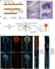
          Reissner’s fiber (RF) is an extracellular polymer comprising the large monomeric protein SCO-spondin (SSPO) secreted by the subcommissural organ (SCO) that extends through cerebrospinal fluid (CSF)-filled ventricles into the central canal of the spinal cord. In zebrafish, RF and CSF-contacting neurons (CSF-cNs) form an axial sensory system that detects spinal curvature, instructs morphogenesis of the body axis, and enables proper alignment of the spine. In mammalian models, RF has been implicated in CSF circulation. However, challenges in manipulating Sspo, an exceptionally large gene of 15,719 nucleotides, with traditional approaches has limited progress. Here, we generated a Sspo knockout mouse model using CRISPR/Cas9-mediated genome-editing. Sspo knockout mice lacked RF-positive material in the SCO and fibrillar condensates in the brain ventricles. Remarkably, Sspo knockout brain ventricle sizes were reduced compared to littermate controls. Minor defects in thoracic spine curvature were detected in Sspo knockouts, which did not alter basic motor behaviors tested. Altogether, our work in mouse demonstrates that SSPO and RF regulate ventricle size during development but only moderately impact spine geometry.

          Supplementary Information

          The online version contains supplementary material available at 10.1186/s12987-023-00491-8.

          Related collections

          Most cited references70

          • Record: found
          • Abstract: found
          • Article: not found

          Fiji: an open-source platform for biological-image analysis.

          Fiji is a distribution of the popular open-source software ImageJ focused on biological-image analysis. Fiji uses modern software engineering practices to combine powerful software libraries with a broad range of scripting languages to enable rapid prototyping of image-processing algorithms. Fiji facilitates the transformation of new algorithms into ImageJ plugins that can be shared with end users through an integrated update system. We propose Fiji as a platform for productive collaboration between computer science and biology research communities.
            Bookmark
            • Record: found
            • Abstract: not found
            • Article: not found

            Fitting Linear Mixed-Effects Models Usinglme4

              Bookmark
              • Record: found
              • Abstract: found
              • Article: not found

              Age-Dependent Niche Signals from the Choroid Plexus Regulate Adult Neural Stem Cells.

              Specialized niches support the lifelong maintenance and function of tissue-specific stem cells. Adult neural stem cells in the ventricular-subventricular zone (V-SVZ) contact the cerebrospinal fluid (CSF), which flows through the lateral ventricles. A largely ignored component of the V-SVZ stem cell niche is the lateral ventricle choroid plexus (LVCP), a primary producer of CSF. Here we show that the LVCP, in addition to performing important homeostatic support functions, secretes factors that promote colony formation and proliferation of purified quiescent and activated V-SVZ stem cells and transit-amplifying cells. The functional effect of the LVCP secretome changes throughout the lifespan, with activated neural stem cells being especially sensitive to age-related changes. Transcriptome analysis identified multiple factors that recruit colony formation and highlights novel facets of LVCP function. Thus, the LVCP is a key niche compartment that translates physiological changes into molecular signals directly affecting neural stem cell behavior.
                Bookmark

                Author and article information

                Contributors
                claire.wyart@icm-institute.org
                maria.lehtinen@childrens.harvard.edu
                Journal
                Fluids Barriers CNS
                Fluids Barriers CNS
                Fluids and Barriers of the CNS
                BioMed Central (London )
                2045-8118
                5 December 2023
                5 December 2023
                2023
                : 20
                : 89
                Affiliations
                [1 ]GRID grid.38142.3c, ISNI 000000041936754X, Department of Pathology, , Boston Children’s Hospital and Harvard Medical School, ; Boston, MA 02115 USA
                [2 ]GRID grid.440907.e, ISNI 0000 0004 1784 3645, Neurophysiology of Brain Circuits, Institut de Biologie de l’Ecole Normale Supérieure (IBENS), Ecole Normale Supérieure, CNRS, INSERM, , Université PSL, ; 75005 Paris, France
                [3 ]GRID grid.50550.35, ISNI 0000 0001 2175 4109, Sorbonne Université, Paris Brain Institute (Institut du Cerveau, ICM), Institut National de la Santé et de la Recherche Médicale (INSERM) U1127, Centre National de la Recherche Scientifique (CNRS) Unité Mixte de Recherche 7225, Assistance Publique–Hôpitaux de Paris (APHP), , Campus Hospitalier Pitié-Salpêtrière, ; 47, bld Hospital, 75013 Paris, France
                Article
                491
                10.1186/s12987-023-00491-8
                10696872
                38049798
                e232dda7-0774-4c2b-8954-bcc85a6ce5b5
                © The Author(s) 2023

                Open Access This article is licensed under a Creative Commons Attribution 4.0 International License, which permits use, sharing, adaptation, distribution and reproduction in any medium or format, as long as you give appropriate credit to the original author(s) and the source, provide a link to the Creative Commons licence, and indicate if changes were made. The images or other third party material in this article are included in the article's Creative Commons licence, unless indicated otherwise in a credit line to the material. If material is not included in the article's Creative Commons licence and your intended use is not permitted by statutory regulation or exceeds the permitted use, you will need to obtain permission directly from the copyright holder. To view a copy of this licence, visit http://creativecommons.org/licenses/by/4.0/. The Creative Commons Public Domain Dedication waiver ( http://creativecommons.org/publicdomain/zero/1.0/) applies to the data made available in this article, unless otherwise stated in a credit line to the data.

                History
                : 1 August 2023
                : 18 November 2023
                Funding
                Funded by: William Randolph Hearst Fund
                Funded by: a Cotrel Spinal Research Foundation
                Funded by: Human Frontier Science Program (HFSP) research program grant
                Award ID: RGP0063/2018
                Award ID: RGP0063/2018
                Award Recipient :
                Funded by: the New York Stem Cell Foundation
                Funded by: the European Research Council
                Award ID: 101002870
                Funded by: FundRef http://dx.doi.org/10.13039/100000002, National Institutes of Health;
                Award ID: NS088566
                Award Recipient :
                Categories
                Research
                Custom metadata
                © BioMed Central Ltd., part of Springer Nature 2023

                Neurology
                sco-spondin/sspo,subcommissural organ (sco),reissner’s fiber,cerebrospinal fluid,spinal cord,ventricular volume,spine deformities

                Comments

                Comment on this article