4
views
0
recommends
+1 Recommend
0 collections
    0
    shares
      • Record: found
      • Abstract: found
      • Article: found
      Is Open Access

      A Step-by-Step Sonographic Approach to Vascular Anomalies in the Pediatric Population: A Pictorial Essay

      research-article

      Read this article at

      Bookmark
          There is no author summary for this article yet. Authors can add summaries to their articles on ScienceOpen to make them more accessible to a non-specialist audience.

          Abstract

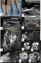
          Vascular anomalies are a common cause of soft-tissue masses in children and often referred for ultrasonographic (USG) evaluation. They are broadly classified as vascular tumors (hemangiomas, hemangioendotheliomas, and angiosarcomas) or vascular malformations (venous malformations, lymphatic malformations, and arteriovenous malformations). Findings on USG and Doppler imaging can be used to categorize vascular anomalies into high- or low-flow lesions, which forms the basis for further workup, diagnosis, and management. On careful evaluation of various sonographic features, in conjunction with clinical findings, an accurate clinicoradiological diagnosis can be made in most cases. Further imaging with magnetic resonance (MR) imaging or computed tomography (CT) helps in delineation of lesion extent, whereas MR or CT angiography is useful to map the vascular supply of high-flow lesions. We have illustrated and discussed a step-by-step approach to diagnose vascular anomalies using ultrasound and Doppler imaging.

          Related collections

          Most cited references39

          • Record: found
          • Abstract: not found
          • Article: not found

          HEMANGIOPERICYTOMA: A VASCULAR TUMOR FEATURING ZIMMERMANN'S PERICYTES.

            Bookmark
            • Record: found
            • Abstract: found
            • Article: not found

            Extracranial arteriovenous malformations: natural progression and recurrence after treatment.

            Arteriovenous malformation is a dynamic vascular anomaly; it expands with age and after treatment. This study analyzed the pattern of arteriovenous malformation progression and frequency of recurrence after therapy.
              Bookmark
              • Record: found
              • Abstract: found
              • Article: not found

              Hemangiomas of infancy.

              Hemangiomas of infancy are unique, benign, pediatric tumors of endothelial cells characterized by an initial phase of rapid proliferation, followed by slow involution, often leading to complete regression. Although most of these tumors are small and innocuous, some may be may be life- or function-threatening, or have associated structural congenital anomalies. Uncertainties regarding their diagnosis or management often prompt referral to a dermatologist. The pathogenesis of hemangiomas of infancy is not well understood, but recent findings suggest a unique vascular phenotype with dysregulated vascular homeostasis. This article reviews new information regarding the pathogenesis of these tumors and highlights the more worrisome presentations, including syndromic hemangiomas, that are likely to be problematic. In addition, management strategies and treatment options are discussed. (J Am Acad Dermatol 2003;48:477-93.) At the completion of this learning activity, participants should be able to describe the clinical features of hemangiomas of infancy and potential complications as well as to understand the strengths and limitations of various treatment options.
                Bookmark

                Author and article information

                Journal
                Indian J Radiol Imaging
                Indian J Radiol Imaging
                10.1055/s-00050590
                The Indian Journal of Radiology & Imaging
                Thieme Medical and Scientific Publishers Private Ltd. (A-12, Second Floor, Sector -2, NOIDA -201301, India )
                0971-3026
                1998-3808
                January 2021
                13 May 2021
                : 31
                : 1
                : 157-171
                Affiliations
                [1 ]Department of Radio-Diagnosis, Lady Hardinge Medical College and Associated Hospitals, New Delhi, India
                [2 ]Department of Pediatric Surgery, Lady Hardinge Medical College and Associated Hospitals, New Delhi, India
                Author notes
                Address for correspondence Pooja Abbey, MD Department of Radio-Diagnosis Lady Hardinge Medical College and Associated Hospitals, New Delhi - 110 001India pooja_abbey@ 123456yahoo.co.in
                Article
                IJRI9520
                10.1055/s-0041-1729486
                8299503
                34316124
                e1e4b101-2a32-4a91-8055-9e673add3ee4
                Indian Radiological Association. This is an open access article published by Thieme under the terms of the Creative Commons Attribution-NonDerivative-NonCommercial-License, permitting copying and reproduction so long as the original work is given appropriate credit. Contents may not be used for commercial purposes, or adapted, remixed, transformed or built upon. (https://creativecommons.org/licenses/by-nc-nd/4.0/).

                This is an open-access article distributed under the terms of the Creative Commons Attribution-NonCommercial-NoDerivatives License, which permits unrestricted reproduction and distribution, for non-commercial purposes only; and use and reproduction, but not distribution, of adapted material for non-commercial purposes only, provided the original work is properly cited.

                History
                Categories
                Pictorial Essay

                Radiology & Imaging
                arteriovenous malformation,doppler,hemangioma,lymphatic malformation,vascular anomalies,vascular malformations,venous malformation

                Comments

                Comment on this article