7
views
0
recommends
+1 Recommend
0 collections
    0
    shares
      • Record: found
      • Abstract: found
      • Article: found
      Is Open Access

      Vascular Occlusions following Ocular Surgical Procedures: A Clinical Observation of Vascular Complications after Ocular Surgery

      research-article

      Read this article at

      Bookmark
          There is no author summary for this article yet. Authors can add summaries to their articles on ScienceOpen to make them more accessible to a non-specialist audience.

          Abstract

          Background

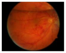
          Ocular vascular occlusions following intraocular procedures are a rare complication. We report a case series of patients with retinal vascular occlusions or anterior ischemic optic neuropathy (AION) after anterior and posterior segment surgery and demonstrate possible risk factors.

          Methods

          Observational case series.

          Results

          In ten patients, vascular occlusions were observed within ten weeks after intraocular surgery: branch retinal arterial occlusion (BRAO) ( n = 2), central retinal artery occlusion (CRAO) ( n = 2), central retinal vein occlusion (CRVO) ( n = 1), branch retinal vein occlusion (BRVO) ( n = 1), anterior ischemic optic neuropathy (AION) ( n = 3), and combined central artery and vein occlusion ( n = 1). AION occurred later (27–69 d) than arterial occlusions (14–60 d) or venous occlusions (1-2 d). In all cases, either specific surgical manipulations or general vascular disorders were identified as risk factors. In addition to general cardiovascular risk factors (arterial hypertension n = 6, diabetes mellitus n = 4), internal workup disclosed bilateral stenosis of the carotid arteries ( n = 1) and myeloproliferative syndrome ( n = 1).

          Conclusion

          Vascular occlusions after surgical ocular procedures seem to be more frequent when cardiovascular diseases coexist. Surgical maneuvers and intra- or postoperative pressure changes may act as a triggering mechanism in patients with underlying systemic cardiovascular disorders. Affected patients should undergo thorough internal examination to identify possible underlying diseases.

          Related collections

          Most cited references25

          • Record: found
          • Abstract: found
          • Article: not found

          Risk factors for central retinal vein occlusion. The Eye Disease Case-Control Study Group.

          S Sperduto (1996)
          To identify possible risk factors for central retinal vein occlusion (CRVO). Between May 1, 1986, and December 31, 1990, 258 patients with CRVO and 1142 controls were identified at five clinical centers. Data were obtained through interviews, clinical examinations, and laboratory analyses of blood specimens. An increased risk of CRVO was found in persons with systemic hypertension, diabetes mellitus, and open-angle glaucoma. Risk of CRVO decreased with increasing levels of physical activity and increasing levels of alcohol consumption. In women, risk of occlusion decreased with use of postmenopausal estrogens and increased with higher erythrocyte sedimentation rates. Cardiovascular disease, electrocardiographic abnormalities, history of treatment of diabetes mellitus, higher blood glucose levels, lower albumin-globulin ratios, and higher alpha-globulin levels were associated with increased risk only for ischemic CRVO. Systemic hypertension was associated with increased risk for ischemic and nonischemic CRVO, but odds ratios were greater for the ischemic type. Our results suggest a cardiovascular risk profile for persons with CRVO, in particular, patients with the ischemic type. The findings reinforce recommendations to diagnose and treat systemic hypertension, advise patients to increase physical exercise, and consider use of exogenous estrogens in postmenopausal women.
            Bookmark
            • Record: found
            • Abstract: found
            • Article: not found

            Management of glaucoma after retinal detachment surgery.

            Secondary glaucoma may complicate retinal detachment surgery. Intraocular pressure (IOP) elevation has been described after scleral buckling procedures and vitrectomy with intravitreal injection of gas or silicone oil. Angle-closure glaucoma after scleral buckling develops because of congestion and anterior rotation of the ciliary body. Medical therapy and laser iridoplasty are usually successful in controlling IOP, but the presence of conjunctival scarring and recession and retinal hardware after scleral buckling procedures can make surgical management challenging. Intravitreal injection of expansile gases like sulfur hexafluoride (SF6) and perfluoropropene (C3F8) may produce secondary angle-closure glaucoma with or without pupillary block. Aspiration of a portion of the intraocular gas may be needed, especially if IOP is elevated to a level that may compromise ocular perfusion. Glaucoma also can develop after intravitreal injection of silicone oil secondary to pupillary block, inflammation, synechial angle closure, rubeosis iridis, or migration of emulsified or nonemulsified silicone oil into the anterior chamber. A prophylactic inferior iridectomy at the time of surgery serves to prevent pupillary block. Patients with medically uncontrolled glaucoma after silicone oil injection may require oil removal with or without concurrent glaucoma surgery.
              Bookmark
              • Record: found
              • Abstract: found
              • Article: not found

              Risk factors for elevated intraocular pressure after the use of intraocular gases in vitreoretinal surgery.

              The authors studied the contribution of multiple factors, including gas type and concentration, to postoperative intraocular pressure (IOP) elevation following vitreoretinal surgery with intraocular gas.
                Bookmark

                Author and article information

                Journal
                J Ophthalmol
                J Ophthalmol
                JOPH
                Journal of Ophthalmology
                Hindawi
                2090-004X
                2090-0058
                2017
                11 July 2017
                : 2017
                : 9120892
                Affiliations
                1University Eye Clinic, Georg-August-Universität Göttingen, Göttingen, Germany
                2Eye Clinic, University Medical Center Schleswig-Holstein, Lübeck, Germany
                3University Eye Clinic, Charité, Berlin, Germany
                4Eye Clinic, University Medical Center Schleswig-Holstein, Kiel, Germany
                Author notes

                Academic Editor: Dirk Sandner

                Author information
                http://orcid.org/0000-0001-9741-9227
                Article
                10.1155/2017/9120892
                5525065
                e1b1ffa0-2703-4d17-8b07-e8cf43e4985e
                Copyright © 2017 Charlotte Fischer et al.

                This is an open access article distributed under the Creative Commons Attribution License, which permits unrestricted use, distribution, and reproduction in any medium, provided the original work is properly cited.

                History
                : 28 March 2017
                : 29 May 2017
                Categories
                Clinical Study

                Ophthalmology & Optometry
                Ophthalmology & Optometry

                Comments

                Comment on this article