0
views
0
recommends
+1 Recommend
0 collections
    0
    shares
      • Record: found
      • Abstract: found
      • Article: found
      Is Open Access

      OSR1 disruption contributes to uterine factor infertility via impaired Müllerian duct development and endometrial receptivity

      research-article

      Read this article at

      Bookmark
          There is no author summary for this article yet. Authors can add summaries to their articles on ScienceOpen to make them more accessible to a non-specialist audience.

          Abstract

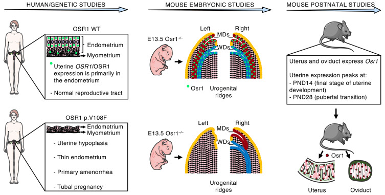
          Three sisters, born from consanguineous parents, manifested a unique Müllerian anomaly characterized by uterine hypoplasia with thin estrogen-unresponsive endometrium and primary amenorrhea, but with spontaneous tubal pregnancies. Through whole-exome sequencing followed by comprehensive genetic analysis, a missense variant was identified in the OSR1 gene. We therefore investigated OSR1/OSR1 expression in postpubertal human uteri, and the prenatal and postnatal expression pattern of Osr1/Osr1 in murine developing Müllerian ducts (MDs) and endometrium, respectively. We then investigated whether Osr1 deletion would affect MD development, using WT and genetically engineered mice. Human uterine OSR1/OSR1 expression was found primarily in the endometrium. Mouse Osr1 was expressed prenatally in MDs and Wolffian ducts (WDs), from rostral to caudal segments, in E13.5 embryos. MDs and WDs were absent on the left side and MDs were rostrally truncated on the right side of E13.5 Osr1 –/– embryos. Postnatally, Osr1 was expressed in mouse uteri throughout their lifespan, peaking at postnatal days 14 and 28. Osr1 protein was present primarily in uterine luminal and glandular epithelial cells and in the epithelial cells of mouse oviducts. Through this translational approach, we demonstrated that OSR1 in humans and mice is important for MD development and endometrial receptivity and may be implicated in uterine factor infertility.

          Abstract

          Related collections

          Most cited references42

          • Record: found
          • Abstract: found
          • Article: not found

          Analysis of relative gene expression data using real-time quantitative PCR and the 2(-Delta Delta C(T)) Method.

          The two most commonly used methods to analyze data from real-time, quantitative PCR experiments are absolute quantification and relative quantification. Absolute quantification determines the input copy number, usually by relating the PCR signal to a standard curve. Relative quantification relates the PCR signal of the target transcript in a treatment group to that of another sample such as an untreated control. The 2(-Delta Delta C(T)) method is a convenient way to analyze the relative changes in gene expression from real-time quantitative PCR experiments. The purpose of this report is to present the derivation, assumptions, and applications of the 2(-Delta Delta C(T)) method. In addition, we present the derivation and applications of two variations of the 2(-Delta Delta C(T)) method that may be useful in the analysis of real-time, quantitative PCR data. Copyright 2001 Elsevier Science (USA).
            Bookmark
            • Record: found
            • Abstract: found
            • Article: found
            Is Open Access

            The mutational constraint spectrum quantified from variation in 141,456 humans

            Genetic variants that inactivate protein-coding genes are a powerful source of information about the phenotypic consequences of gene disruption: genes that are crucial for the function of an organism will be depleted of such variants in natural populations, whereas non-essential genes will tolerate their accumulation. However, predicted loss-of-function variants are enriched for annotation errors, and tend to be found at extremely low frequencies, so their analysis requires careful variant annotation and very large sample sizes 1 . Here we describe the aggregation of 125,748 exomes and 15,708 genomes from human sequencing studies into the Genome Aggregation Database (gnomAD). We identify 443,769 high-confidence predicted loss-of-function variants in this cohort after filtering for artefacts caused by sequencing and annotation errors. Using an improved model of human mutation rates, we classify human protein-coding genes along a spectrum that represents tolerance to inactivation, validate this classification using data from model organisms and engineered human cells, and show that it can be used to improve the power of gene discovery for both common and rare diseases.
              Bookmark
              • Record: found
              • Abstract: found
              • Article: not found

              From FastQ data to high confidence variant calls: the Genome Analysis Toolkit best practices pipeline.

              This unit describes how to use BWA and the Genome Analysis Toolkit (GATK) to map genome sequencing data to a reference and produce high-quality variant calls that can be used in downstream analyses. The complete workflow includes the core NGS data processing steps that are necessary to make the raw data suitable for analysis by the GATK, as well as the key methods involved in variant discovery using the GATK.
                Bookmark

                Author and article information

                Contributors
                Journal
                J Clin Invest
                J Clin Invest
                J Clin Invest
                The Journal of Clinical Investigation
                American Society for Clinical Investigation
                0021-9738
                1558-8238
                1 December 2023
                1 December 2023
                1 December 2023
                : 133
                : 23
                : e161701
                Affiliations
                [1 ]Molecular Pharmacology Laboratory (FARMOL), Faculty of Health Sciences, University of Brasilia, Brasilia-DF, Brazil.
                [2 ]Section of Endocrinology, Gonadal and Adrenal Diseases Clinics, University Hospital of Brasilia, Brasilia-DF, Brazil.
                [3 ]Division of Endocrinology, Diabetes and Hypertension, Brigham and Women’s Hospital and Harvard Medical School, Boston, Massachusetts, USA.
                [4 ]Division of Endocrinology, Children’s National Hospital, Washington, DC, USA.
                [5 ]Department of Pediatrics, George Washington School of Medicine and Health Sciences, Washington, DC, USA.
                [6 ]Whitehead Institute for Biomedical Research, Cambridge, Massachusetts, USA.
                [7 ]Division of Endocrinology, Boston Children’s Hospital, Boston, Massachusetts, USA.
                [8 ]Program in Medical and Population Genetics, Broad Institute, Cambridge, Massachusetts, USA.
                [9 ]Department of Genetics, Harvard Medical School, Boston, Massachusetts, USA.
                [10 ]Department of Genetics and Morphology, Institute of Biology, University of Brasilia, Brasilia-DF, Brazil.
                [11 ]Jackson Laboratory for Genomic Medicine, Farmington, Connecticut, USA.
                [12 ]Division of Developmental Biology, Cincinnati Children’s Hospital Medical Center, Cincinnati, Ohio, USA.
                [13 ]Department of Obstetrics and Gynecology, Boston University Chobanian & Avedisian School of Medicine, Boston, Massachusetts, USA.
                [14 ]Department of Pediatrics, University of Cincinnati College of Medicine, Cincinnati, Ohio, USA.
                Author notes
                Address correspondence to: Ursula B. Kaiser, Division of Endocrinology, Diabetes and Hypertension, Brigham and Women’s Hospital, 221 Longwood Avenue, Boston Massachusetts 02115, USA. Phone: 1.617.525.8867; Email: ukaiser@ 123456bwh.harvard.edu .

                Authorship note: ALP and SAP contributed equally to this work.

                Author information
                http://orcid.org/0000-0002-7937-2952
                http://orcid.org/0000-0002-5989-9255
                http://orcid.org/0000-0002-9292-6024
                http://orcid.org/0000-0002-5505-0852
                http://orcid.org/0000-0002-7741-0257
                http://orcid.org/0000-0001-7842-4696
                http://orcid.org/0000-0002-8237-0704
                Article
                161701
                10.1172/JCI161701
                10688984
                37847567
                e14c9b16-b04b-48d7-924d-f508fd5ee520
                © 2023 Lofrano-Porto et al.

                This work is licensed under the Creative Commons Attribution 4.0 International License. To view a copy of this license, visit http://creativecommons.org/licenses/by/4.0/.

                History
                : 13 May 2022
                : 28 September 2023
                Funding
                Funded by: National Institutes of Health (NIH)
                Award ID: DE013681,R37HD019938,R01HD082314,R21HD098684
                Funded by: Brazilian National Council for Scientific and Technological Development (CNPq)
                Award ID: PDE 208395/2014-8,MCTI/CNPq/Universal 462346/2014-5
                Funded by: Coordination of Superior Level Staff Improvement (CAPES)
                Award ID: PDSE 88881.135648/2016-01
                Categories
                Research Article

                development,reproductive biology,embryonic development,genetic diseases,obstetrics/gynecology

                Comments

                Comment on this article