0
views
0
recommends
+1 Recommend
0 collections
    0
    shares
      • Record: found
      • Abstract: found
      • Article: found
      Is Open Access

      Bacterial outer membrane vesicle based versatile nanosystem boosts the efferocytosis blockade triggered tumor-specific immunity

      research-article

      Read this article at

      Bookmark
          There is no author summary for this article yet. Authors can add summaries to their articles on ScienceOpen to make them more accessible to a non-specialist audience.

          Abstract

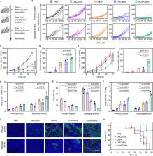
          Efferocytosis inhibition is emerging as an attractive strategy for antitumor immune therapy because of the subsequent leak of abundant immunogenic contents. However, the practical efficacy is seriously impeded by the immunosuppressive tumor microenvironments. Here, we construct a versatile nanosystem that can not only inhibit the efferocytosis but also boost the following antitumor immunity. MerTK inhibitor UNC2025 is loaded into the bacterial outer membrane vesicles (OMVs), which are then modified with maleimide (mU@OMVs). The prepared mU@OMVs effectively inhibits the efferocytosis by promoting the uptake while preventing the MerTK phosphorylation of tumor associated macrophages, and then captures the released antigens through forming universal thioether bonds. The obtained in situ vaccine effectively transfers to lymph nodes by virtue of the intrinsic features of OMVs, and then provokes intense immune responses that can efficiently prevent the growth, metastasis and recurrence of tumors in mice, providing a generalizable strategy for cancer immunotherapy.

          Abstract

          Efferocytosis inhibition leads to the release of immunogenic contents into the tumor microenvironment. Here the authors developed a nanosystem that inhibits MerTK-mediated efferocytosis and captures tumor-associated agents to enhance antitumour immunity.

          Related collections

          Most cited references47

          • Record: found
          • Abstract: found
          • Article: not found

          Release of chromatin protein HMGB1 by necrotic cells triggers inflammation.

          High mobility group 1 (HMGB1) protein is both a nuclear factor and a secreted protein. In the cell nucleus it acts as an architectural chromatin-binding factor that bends DNA and promotes protein assembly on specific DNA targets. Outside the cell, it binds with high affinity to RAGE (the receptor for advanced glycation end products) and is a potent mediator of inflammation. HMGB1 is secreted by activated monocytes and macrophages, and is passively released by necrotic or damaged cells. Here we report that Hmgb1(-/-) necrotic cells have a greatly reduced ability to promote inflammation, which proves that the release of HMGB1 can signal the demise of a cell to its neighbours. Apoptotic cells do not release HMGB1 even after undergoing secondary necrosis and partial autolysis, and thus fail to promote inflammation even if not cleared promptly by phagocytic cells. In apoptotic cells, HMGB1 is bound firmly to chromatin because of generalized underacetylation of histone and is released in the extracellular medium (promoting inflammation) if chromatin deacetylation is prevented. Thus, cells undergoing apoptosis are programmed to withhold the signal that is broadcast by cells that have been damaged or killed by trauma.
            Bookmark
            • Record: found
            • Abstract: found
            • Article: not found

            Using immunotherapy to boost the abscopal effect

            More than 60 years ago, the effect whereby radiotherapy at one site may lead to regression of metastatic cancer at distant sites that are not irradiated was described and called the abscopal effect (from ‘ab scopus’, that is, away from the target). The abscopal effect has been connected to mechanisms involving the immune system. However, the effect is rare because at the time of treatment, established immune-tolerance mechanisms may hamper the development of sufficiently robust abscopal responses. Today, the growing consensus is that combining radiotherapy with immunotherapy provides an opportunity to boost abscopal response rates, extending the use of radiotherapy to treatment of both local and metastatic disease. In this Opinion article, we review evidence for this growing consensus and highlight emerging limitations to boosting the abscopal effect using immunotherapy. This is followed by a perspective on current and potential cross-disciplinary approaches, including the use of smart materials to address these limitations.
              Bookmark
              • Record: found
              • Abstract: found
              • Article: not found

              Cd8+ but Not Cd8− Dendritic Cells Cross-Prime Cytotoxic T Cells in Vivo

              Bone marrow–derived antigen-presenting cells (APCs) take up cell-associated antigens and present them in the context of major histocompatibility complex (MHC) class I molecules to CD8+ T cells in a process referred to as cross-priming. Cross-priming is essential for the induction of CD8+ T cell responses directed towards antigens not expressed in professional APCs. Although in vitro experiments have shown that dendritic cells (DCs) and macrophages are capable of presenting exogenous antigens in association with MHC class I, the cross-presenting cell in vivo has not been identified. We have isolated splenic DCs after in vivo priming with ovalbumin-loaded β2-microglobulin–deficient splenocytes and show that they indeed present cell-associated antigens in the context of MHC class I molecules. This process is transporter associated with antigen presentation (TAP) dependent, suggesting an endosome to cytosol transport. To determine whether a specific subset of splenic DCs is involved in this cross-presentation, we negatively and positively selected for CD8− and CD8+ DCs. Only the CD8+, and not the CD8−, DC subset demonstrates cross-priming ability. FACS® studies after injection of splenocytes loaded with fluorescent beads showed that 1 and 0.6% of the CD8+ and the CD8− DC subsets, respectively, had one or more associated beads. These results indicate that CD8+ DCs play an important role in the generation of cytotoxic T lymphocyte responses specific for cell-associated antigens.
                Bookmark

                Author and article information

                Contributors
                hyanxie@bit.edu.cn
                Journal
                Nat Commun
                Nat Commun
                Nature Communications
                Nature Publishing Group UK (London )
                2041-1723
                25 March 2023
                25 March 2023
                2023
                : 14
                : 1675
                Affiliations
                [1 ]GRID grid.43555.32, ISNI 0000 0000 8841 6246, School of Life Science, , Beijing Institute of Technology, ; 100081 Beijing, P.R. China
                [2 ]GRID grid.43555.32, ISNI 0000 0000 8841 6246, School of Medical Technology, , Beijing Institute of Technology, ; 100081 Beijing, P.R. China
                Author information
                http://orcid.org/0000-0002-6330-7929
                Article
                37369
                10.1038/s41467-023-37369-0
                10039929
                36966130
                e0711d28-03e5-401d-a346-12beee33573c
                © The Author(s) 2023

                Open Access This article is licensed under a Creative Commons Attribution 4.0 International License, which permits use, sharing, adaptation, distribution and reproduction in any medium or format, as long as you give appropriate credit to the original author(s) and the source, provide a link to the Creative Commons license, and indicate if changes were made. The images or other third party material in this article are included in the article’s Creative Commons license, unless indicated otherwise in a credit line to the material. If material is not included in the article’s Creative Commons license and your intended use is not permitted by statutory regulation or exceeds the permitted use, you will need to obtain permission directly from the copyright holder. To view a copy of this license, visit http://creativecommons.org/licenses/by/4.0/.

                History
                : 22 August 2022
                : 14 March 2023
                Funding
                Funded by: FundRef https://doi.org/10.13039/501100001809, National Natural Science Foundation of China (National Science Foundation of China);
                Award ID: 21874011, 21904012, 91859123, 32101140, 22104005
                Award Recipient :
                Funded by: FundRef https://doi.org/10.13039/501100002858, China Postdoctoral Science Foundation;
                Award ID: 2021TQ0037, 2021M690405
                Award Recipient :
                Funded by: National Science Fund for Distinguished Young Scholars (22025401) Beijing Institute of Technology Research Fund Program for Young Scholars (XSQD-202212002)
                Categories
                Article
                Custom metadata
                © The Author(s) 2023

                Uncategorized
                tumour vaccines,cancer immunotherapy
                Uncategorized
                tumour vaccines, cancer immunotherapy

                Comments

                Comment on this article