42
views
0
recommends
+1 Recommend
0 collections
    0
    shares
      • Record: found
      • Abstract: found
      • Article: found
      Is Open Access

      Ossicle in Anterior Cruciate Ligament: A Rare Occurrence

      case-report

      Read this article at

      Bookmark
          There is no author summary for this article yet. Authors can add summaries to their articles on ScienceOpen to make them more accessible to a non-specialist audience.

          Abstract

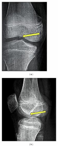
          The occurrence of an intra-articular ossicle is not rare in the knee, with reports suggesting the existence of meniscal osscile. There are also reports describing the attachment of the posterolateral bundle of the anterior cruciate ligament (ACL) to an accessory ossicle. However, despite an extensive search of the English literature we did not find much written about an intrasubstance ossicle in the ACL. We present the case of a 13-year-old male with an intrasubstance ossicle in the anteromedial bundle of the ACL of his right knee.

          Related collections

          Most cited references14

          • Record: found
          • Abstract: found
          • Article: not found

          Intraarticular ganglia of the knee: prevalence, presentation, etiology, and management.

          The aim of this study was to present a comprehensive review of intraarticular ganglia of the knee. Cases of intraarticular ganglia of the knee were collected from a group of 1767 consecutive patients referred for MR imaging examinations of the knee. Medical records and radiologic, arthroscopic, and pathologic reports were reviewed. Twenty-three patients (17 men and six women) had intraarticular ganglia of the knee. Three ganglia were found in Hoffa's fat pad, 10 were associated with the anterior cruciate ligament, eight were associated with the posterior cruciate ligament, and two had no definite association with either the anterior or the posterior cruciate ligament. Of the 23 ganglia, 14 (61%) were located in the intercondylar notch. Pain was the most common complaint: medial joint line tenderness, lateral joint line tenderness, or retropatellar pain. Two of the three patients with ganglia in Hoffa's fat pad had a palpable mass on physical examination. Of the 23 patients with intraarticular ganglia of the knee, 18 (78%) had no associated internal derangement. Five patients underwent arthroscopic surgery, confirming the diagnosis of ganglionic cyst. Of these five patients, none had a recurrence. With the advent of MR imaging of the knee, intraarticular ganglia of the knee have been found to occur more commonly than once thought, with a prevalence of 1.3% in this series. Radiologists interpreting knee MR images must be aware of this entity and its management.
            Bookmark
            • Record: found
            • Abstract: found
            • Article: not found

            Meniscal ossicle: radiographic and MR imaging findings.

            To determine the magnetic resonance (MR) imaging appearance of meniscal ossicles and correlate that with the radiographic and clinical features. Radiographs and MR images were studied retrospectively in six patients with meniscal ossicles; all patients had intermittent discomfort in the knee, and five patients had experienced knee trauma. Imaging findings were correlated with the clinical history. Four patients underwent arthroscopy. All patients had a visible ossicle on conventional radiographs initially interpreted as a loose joint body. MR imaging features included a corticated marrow-containing structure within the substance of the medial meniscus near the tibial attachment that appeared as a rounded focus of increased signal intensity on T1-weighted images that decreased in signal intensity on T2-weighted images. Meniscal ossicles have a characteristic MR appearance that may help distinguish them from loose bodies. They should be considered diagnostically when a circumscribed ossification is identified near the posterior horn of the medial meniscus on radiographs.
              Bookmark
              • Record: found
              • Abstract: found
              • Article: not found

              Symptomatic calcification of the anterior cruciate ligament: A case report.

              We report a rare case of symptomatic calcification of the ACL. A 31-year-old man complained of severe knee pain with restriction of knee motion from 30° to 130° for a week. Plain radiographs and multi-planar CT revealed calcification within the intercondylar notch with no osteoarthritic changes. MRI revealed a low signal intensity mass near the intact ACL. The ACL appeared bulged by arthroscopy and white and creamy fluid exuded from the partially excised synovial membrane. Pain subsided immediately postoperatively. Histologically, the calcific deposit near the ACL showed negligible degenerative changes and resembled calcifying tendinitis of the rotator cuff. Although calcific deposits rarely affect the knee joint, calcification of the ACL should be included in differential diagnoses for acute knee pain and restricted range of motion such as mechanical locking. This case illustrates that arthroscopic removal of the deposits can be effective.
                Bookmark

                Author and article information

                Journal
                Case Rep Orthop
                Case Rep Orthop
                CRIOR
                Case Reports in Orthopedics
                Hindawi Publishing Corporation
                2090-6749
                2090-6757
                2014
                13 April 2014
                : 2014
                : 616715
                Affiliations
                Department of Orthopaedics, Pt. B.D. Sharma PGIMS, Rohatk, Haryana 124001, India
                Author notes
                *Reetadyuti Mukhopadhyay: reetya_m@ 123456hotmail.com

                Academic Editor: John Nyland

                Author information
                http://orcid.org/0000-0002-0475-7145
                http://orcid.org/0000-0002-8347-4943
                http://orcid.org/0000-0002-3043-7129
                http://orcid.org/0000-0003-4771-4636
                Article
                10.1155/2014/616715
                4009161
                24822144
                e0705338-e1a9-4b52-8a25-f236edfa928c
                Copyright © 2014 Ashish Devgan et al.

                This is an open access article distributed under the Creative Commons Attribution License, which permits unrestricted use, distribution, and reproduction in any medium, provided the original work is properly cited.

                History
                : 18 January 2014
                : 17 March 2014
                : 28 March 2014
                Categories
                Case Report

                Orthopedics
                Orthopedics

                Comments

                Comment on this article