12
views
0
recommends
+1 Recommend
0 collections
    0
    shares
      • Record: found
      • Abstract: found
      • Article: found
      Is Open Access

      Bone Marrow CD133 + Stem Cells Ameliorate Visual Dysfunction in Streptozotocin-induced Diabetic Mice with Early Diabetic Retinopathy

      research-article

      Read this article at

      Bookmark
          There is no author summary for this article yet. Authors can add summaries to their articles on ScienceOpen to make them more accessible to a non-specialist audience.

          Abstract

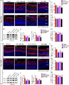
          Diabetic retinopathy (DR), one of the leading causes of vision loss worldwide, is characterized by neurovascular disorders. Emerging evidence has demonstrated retinal neurodegeneration in the early pathogenesis of DR, and no treatment has been developed to prevent the early neurodegenerative changes that precede detectable microvascular disorders. Bone marrow CD133 + stem cells with revascularization properties exhibit neuroregenerative potential. However, whether CD133 + cells can ameliorate the neurodegeneration at the early stage of DR remains unclear. In this study, mouse bone marrow CD133 + stem cells were immunomagnetically isolated and analyzed for the phenotypic characteristics, capacity for neural differentiation, and gene expression of neurotrophic factors. After being labeled with enhanced green fluorescent protein, CD133 + cells were intravitreally transplanted into streptozotocin (STZ)-induced diabetic mice to assess the outcomes of visual function and retina structure and the mechanism underlying the therapeutic effect. We found that CD133 + cells co-expressed typical hematopoietic/endothelial stem/progenitor phenotypes, could differentiate to neural lineage cells, and expressed genes of robust neurotrophic factors in vitro. Functional analysis demonstrated that the transplantation of CD133 + cells prevented visual dysfunction for 56 days. Histological analysis confirmed such a functional improvement and showed that transplanted CD133 + cells survived, migrated into the inner retina (IR) over time and preserved IR degeneration, including retina ganglion cells (RGCs) and rod-on bipolar cells. In addition, a subset of transplanted CD133 + cells in the ganglion cell layer differentiated to express RGC markers in STZ-induced diabetic retina. Moreover, transplanted CD133 + cells expressed brain-derived neurotrophic factors (BDNFs) in vivo and increased the BDNF level in STZ-induced diabetic retina to support the survival of retinal cells. Based on these findings, we suggest that transplantation of bone marrow CD133 + stem cells represents a novel approach to ameliorate visual dysfunction and the underlying IR neurodegeneration at the early stage of DR.

          Related collections

          Most cited references59

          • Record: found
          • Abstract: found
          • Article: found
          Is Open Access

          CD133: a stem cell biomarker and beyond

          Zhong Li (2013)
          Cancer stem cells (CSCs) or tumor initiating cells (TICs) contribute to tumorigenesis, metastasis, recurrence and chemoresistance. CD133, a pentaspan membrane glycoprotein, has been used as a stem cell biomarker for isolation of stem-like cells from a variety of normal and pathological tissues as well as cell lines since its discovery in 1999. Recent studies are focusing on the functionality of CD133. In this review, we summarize new insights into CD133 regulation and the involvement of CD133 in cell self-renewal, tumorigenesis, metastasis, resistance, metabolism, differentiation, autophagy, apoptosis, and regeneration.
            Bookmark
            • Record: found
            • Abstract: found
            • Article: not found

            Diabetes impairs hematopoietic stem cell mobilization by altering niche function.

            Success with transplantation of autologous hematopoietic stem and progenitor cells (HSPCs) in patients depends on adequate collection of these cells after mobilization from the bone marrow niche by the cytokine granulocyte colony-stimulating factor (G-CSF). However, some patients fail to achieve sufficient HSPC mobilization. Retrospective analysis of bone marrow transplant patient records revealed that diabetes correlated with poor mobilization of CD34+ HSPCs. In mouse models of type 1 and type 2 diabetes (streptozotocin-induced and db/db mice, respectively), we found impaired egress of murine HSPCs from the bone marrow after G-CSF treatment. Furthermore, HSPCs were aberrantly localized in the marrow niche of the diabetic mice, and abnormalities in the number and function of sympathetic nerve termini were associated with this mislocalization. Aberrant responses to β-adrenergic stimulation of the bone marrow included an inability of marrow mesenchymal stem cells expressing the marker nestin to down-modulate the chemokine CXCL12 in response to G-CSF treatment (mesenchymal stem cells are reported to be critical for HSPC mobilization). The HSPC mobilization defect was rescued by direct pharmacological inhibition of the interaction of CXCL12 with its receptor CXCR4 using the drug AMD3100. These data suggest that there are diabetes-induced changes in bone marrow physiology and microanatomy and point to a potential intervention to overcome poor HSPC mobilization in diabetic patients.
              Bookmark
              • Record: found
              • Abstract: found
              • Article: not found

              Human CD133+ progenitor cells promote the healing of diabetic ischemic ulcers by paracrine stimulation of angiogenesis and activation of Wnt signaling.

              We evaluated the healing potential of human fetal aorta-derived CD133(+) progenitor cells and their conditioned medium (CD133(+) CCM) in a new model of ischemic diabetic ulcer. Streptozotocin-induced diabetic mice underwent bilateral limb ischemia and wounding. One wound was covered with collagen containing 2x10(4) CD133(+) or CD133(-) cells or vehicle. The contralateral wound, covered with only collagen, served as control. Fetal CD133(+) cells expressed high levels of wingless (Wnt) genes, which were downregulated following differentiation into CD133(-) cells along with upregulation of Wnt antagonists secreted frizzled-related protein (sFRP)-1, -3, and -4. CD133(+) cells accelerated wound closure as compared with CD133(-) or vehicle and promoted angiogenesis through stimulation of endothelial cell proliferation, migration, and survival by paracrine effects. CD133(+) cells secreted high levels of vascular endothelial growth factor (VEGF)-A and interleukin (IL)-8. Consistently, CD133(+) CCM accelerated wound closure and reparative angiogenesis, with this action abrogated by co-administering the Wnt antagonist sFRP-1 or neutralizing antibodies against VEGF-A or IL-8. In vitro, these effects were recapitulated following exposure of high-glucose-primed human umbilical vein endothelial cells to CD133(+) CCM, resulting in stimulation of migration, angiogenesis-like network formation and induction of Wnt expression. The promigratory and proangiogenic effect of CD133(+) CCM was blunted by sFRP-1, as well as antibodies against VEGF-A or IL-8. CD133(+) cells stimulate wound healing by paracrine mechanisms that activate Wnt signaling pathway in recipients. These preclinical findings open new perspectives for the cure of diabetic ulcers.
                Bookmark

                Author and article information

                Journal
                Cell Transplant
                Cell Transplant
                CLL
                spcll
                Cell Transplantation
                SAGE Publications (Sage CA: Los Angeles, CA )
                0963-6897
                1555-3892
                02 May 2018
                June 2018
                : 27
                : 6
                : 916-936
                Affiliations
                [1 ]Southwest Hospital, Southwest Eye Hospital, Third Military Medical University (Army Medical University), Chongqing, China
                [2 ]Key Lab of Visual Damage and Regeneration & Restoration of Chongqing, Chongqing, China
                Author notes
                [*]Haiwei Xu, Southwest Hospital, Southwest Eye Hospital, Third Military Medical University (Army Medical University), 30 Gaotanyan ST, Shapingba District, Chongqing, 400038, China. Email: haiweixu2001@ 123456163.com
                [*]Zheng Qin Yin, Southwest Hospital, Southwest Eye Hospital, Third Military Medical University (Army Medical University), 30 Gaotanyan ST, Shapingba District, Chongqing, 400038, China. Email: qinzyin@ 123456aliyun.com
                Author information
                http://orcid.org/0000-0002-8840-7918
                Article
                10.1177_0963689718759463
                10.1177/0963689718759463
                6050916
                29717657
                e01df26a-9ef1-4c6b-b8f5-0817bc230252
                © The Author(s) 2018

                This article is distributed under the terms of the Creative Commons Attribution-NonCommercial 4.0 License ( http://www.creativecommons.org/licenses/by-nc/4.0/) which permits non-commercial use, reproduction and distribution of the work without further permission provided the original work is attributed as specified on the SAGE and Open Access pages ( https://us.sagepub.com/en-us/nam/open-access-at-sage).

                History
                : 14 December 2017
                : 15 January 2018
                : 22 January 2018
                Funding
                Funded by: National Key Basic Research Program of China;
                Award ID: 973 Project 2013CB967002
                Categories
                Original Articles

                diabetic retinopathy,cd133+ cells,visual function,retina degeneration,cell transplantation

                Comments

                Comment on this article