23
views
0
recommends
+1 Recommend
0 collections
    0
    shares
      • Record: found
      • Abstract: found
      • Article: not found

      Cancer: An Oxidative Crosstalk between Solid Tumor Cells and Cancer Associated Fibroblasts

      review-article

      Read this article at

      ScienceOpenPublisherPMC
      Bookmark
          There is no author summary for this article yet. Authors can add summaries to their articles on ScienceOpen to make them more accessible to a non-specialist audience.

          Abstract

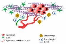
          Redox balance is associated with the regulation of several cell signalling pathways and functions. In fact, under physiological conditions, cells maintain a balance between oxidant and antioxidant systems, and reactive oxygen species (ROS) can act as second messengers to regulate cell proliferation, cell death, and other physiological processes. Cancer tissues usually contain higher levels of ROS than normal tissues, and this ROS overproduction is associated with tumor development. Neoplastic tissues are very heterogeneous systems, composed of tumor cells and microenvironment that has a critical role in tumor progression. Cancer associated fibroblasts (CAFs) represent the main cell type of tumor microenvironment, and they contribute to tumor growth by undergoing an irreversible activation process. It is known that ROS can be transferred from cancer cells to fibroblasts. In particular, ROS affect the behaviour of CAFs by promoting the conversion of fibroblasts to myofibroblasts that support tumor progression and dissemination. Furthermore, the wrecking of redox homeostasis in cancer cells and tumor microenvironment induces a metabolic reprogramming in tumor cells and cancer associated fibroblasts, giving advantage to cancer growth. This review describes the role of ROS in tumor growth, by focusing on CAFs activation and metabolic interactions between cancer cells and stromal fibroblasts.

          Related collections

          Most cited references68

          • Record: found
          • Abstract: found
          • Article: not found

          Specific aquaporins facilitate the diffusion of hydrogen peroxide across membranes.

          The metabolism of aerobic organisms continuously produces reactive oxygen species. Although potentially toxic, these compounds also function in signaling. One important feature of signaling compounds is their ability to move between different compartments, e.g. to cross membranes. Here we present evidence that aquaporins can channel hydrogen peroxide (H2O2). Twenty-four aquaporins from plants and mammals were screened in five yeast strains differing in sensitivity toward oxidative stress. Expression of human AQP8 and plant Arabidopsis TIP1;1 and TIP1;2 in yeast decreased growth and survival in the presence of H2O2. Further evidence for aquaporin-mediated H2O2 diffusion was obtained by a fluorescence assay with intact yeast cells using an intracellular reactive oxygen species-sensitive fluorescent dye. Application of silver ions (Ag+), which block aquaporin-mediated water diffusion in a fast kinetics swelling assay, also reversed both the aquaporin-dependent growth repression and the H2O2-induced fluorescence. Our results present the first molecular genetic evidence for the diffusion of H2O2 through specific members of the aquaporin family.
            Bookmark
            • Record: found
            • Abstract: found
            • Article: found
            Is Open Access

            Cancer metastases: challenges and opportunities

            Cancer metastasis is the major cause of cancer morbidity and mortality, and accounts for about 90% of cancer deaths. Although cancer survival rate has been significantly improved over the years, the improvement is primarily due to early diagnosis and cancer growth inhibition. Limited progress has been made in the treatment of cancer metastasis due to various factors. Current treatments for cancer metastasis are mainly chemotherapy and radiotherapy, though the new generation anti-cancer drugs (predominantly neutralizing antibodies for growth factors and small molecule kinase inhibitors) do have the effects on cancer metastasis in addition to their effects on cancer growth. Cancer metastasis begins with detachment of metastatic cells from the primary tumor, travel of the cells to different sites through blood/lymphatic vessels, settlement and growth of the cells at a distal site. During the process, metastatic cells go through detachment, migration, invasion and adhesion. These four essential, metastatic steps are inter-related and affected by multi-biochemical events and parameters. Additionally, it is known that tumor microenvironment (such as extracellular matrix structure, growth factors, chemokines, matrix metalloproteinases) plays a significant role in cancer metastasis. The biochemical events and parameters involved in the metastatic process and tumor microenvironment have been targeted or can be potential targets for metastasis prevention and inhibition. This review provides an overview of these metastasis essential steps, related biochemical factors, and targets for intervention.
              Bookmark
              • Record: found
              • Abstract: found
              • Article: not found

              Autocrine TGF-beta and stromal cell-derived factor-1 (SDF-1) signaling drives the evolution of tumor-promoting mammary stromal myofibroblasts.

              Much interest is currently focused on the emerging role of tumor-stroma interactions essential for supporting tumor progression. Carcinoma-associated fibroblasts (CAFs), frequently present in the stroma of human breast carcinomas, include a large number of myofibroblasts, a hallmark of activated fibroblasts. These fibroblasts have an ability to substantially promote tumorigenesis. However, the precise cellular origins of CAFs and the molecular mechanisms by which these cells evolve into tumor-promoting myofibroblasts remain unclear. Using a coimplantation breast tumor xenograft model, we show that resident human mammary fibroblasts progressively convert into CAF myofibroblasts during the course of tumor progression. These cells increasingly acquire two autocrine signaling loops, mediated by TGF-β and SDF-1 cytokines, which both act in autostimulatory and cross-communicating fashions. These autocrine-signaling loops initiate and maintain the differentiation of fibroblasts into myofibroblasts and the concurrent tumor-promoting phenotype. Collectively, these findings indicate that the establishment of the self-sustaining TGF-β and SDF-1 autocrine signaling gives rise to tumor-promoting CAF myofibroblasts during tumor progression. This autocrine-signaling mechanism may prove to be an attractive therapeutic target to block the evolution of tumor-promoting CAFs.
                Bookmark

                Author and article information

                Journal
                Biomed Res Int
                Biomed Res Int
                BMRI
                BioMed Research International
                Hindawi Publishing Corporation
                2314-6133
                2314-6141
                2016
                9 August 2016
                : 2016
                : 4502846
                Affiliations
                1Department of Public Health, University of Naples Federico II, 80131 Naples, Italy
                2Department of Molecular Medicine and Medical Biotechnology, University of Naples Federico II, 80131 Naples, Italy
                Author notes
                *Stefania Montagnani: montagna@ 123456unina.it

                Academic Editor: Amit Budhraja

                Author information
                http://orcid.org/0000-0002-7420-372X
                Article
                10.1155/2016/4502846
                4993917
                27595103
                df8a35aa-7f47-4207-ad33-a055e69e2819
                Copyright © 2016 Alessandro Arcucci et al.

                This is an open access article distributed under the Creative Commons Attribution License, which permits unrestricted use, distribution, and reproduction in any medium, provided the original work is properly cited.

                History
                : 27 May 2016
                : 13 July 2016
                Funding
                Funded by: Regione Campania (Italy)
                Award ID: GR-2009-1580434
                Categories
                Review Article

                Comments

                Comment on this article