19
views
0
recommends
+1 Recommend
0 collections
    0
    shares
      • Record: found
      • Abstract: found
      • Article: found
      Is Open Access

      First-in-Humans Evaluation of Safety and Dosimetry of 64Cu-LLP2A for PET Imaging

      research-article

      Read this article at

      Bookmark
          There is no author summary for this article yet. Authors can add summaries to their articles on ScienceOpen to make them more accessible to a non-specialist audience.

          Visual Abstract

          Abstract

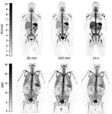
          There remains an unmet need for molecularly targeted imaging agents for multiple myeloma (MM). The integrin very late antigen 4 (VLA4), is differentially expressed in malignant MM cells and in pathogenic inflammatory microenvironmental cells. [ 64Cu]Cu-CB-TE1A1P-LLP2A ( 64Cu-LLP2A) is a VLA4-targeted, high-affinity radiopharmaceutical with promising utility for managing patients diagnosed with MM. Here, we evaluated the safety and human radiation dosimetry of 64Cu-LLP2A for potential use in MM patients. Methods: A single-dose [ natCu]Cu-LLP2A (Cu-LLP2A) tolerability and toxicity study was performed on CD-1 (Hsd:ICR) male and female mice. 64Cu-LLP2A was synthesized in accordance with good-manufacturing-practice–compliant procedures. Three MM patients and six healthy participants underwent 64Cu-LLP2A-PET/CT or PET/MRI at up to 3 time points to help determine tracer biodistribution, pharmacokinetics, and radiation dosimetry. Time–activity curves were plotted for each participant. Mean organ-absorbed doses and effective doses were calculated using the OLINDA software. Tracer bioactivity was evaluated via cell-binding assays, and metabolites from human blood samples were analyzed with analytic radio–high-performance liquid chromatography. When feasible, VLA4 expression was evaluated in the biopsy tissues using 14-color flow cytometry. Results: A 150-fold mass excess of the desired imaging dose was tolerated well in male and female CD-1 mice (no observed adverse effect level). Time–activity curves from human imaging data showed rapid tracer clearance from blood via the kidneys and bladder. The effective dose of 64Cu-LLP2A in humans was 0.036 ± 0.006 mSv/MBq, and the spleen had the highest organ uptake, 0.142 ± 0.034 mSv/MBq. Among all tissues, the red marrow demonstrated the highest residence time. Image quality analysis supports an early imaging time (4–5 h after injection of the radiotracer) as optimal. Cell studies showed statistically significant blocking for the tracer produced for all human studies (82.42% ± 13.47%). Blood metabolism studies confirmed a stable product peak (>90%) up to 1 h after injection of the radiopharmaceutical. No clinical or laboratory adverse events related to 64Cu-LLP2A were observed in the human participants. Conclusion: 64Cu-LLP2A exhibited a favorable dosimetry and safety profile for use in humans.

          Related collections

          Most cited references39

          • Record: found
          • Abstract: found
          • Article: not found

          Feasibility of blood testing combined with PET-CT to screen for cancer and guide intervention

          Cancer treatments are often more successful when the disease is detected early. We evaluated the feasibility and safety of multi-cancer blood testing coupled with PET-CT imaging to detect cancer in a prospective, interventional study of 10,006 women not previously known to have cancer. Positive blood tests were independently confirmed by a diagnostic PET-CT, which also localized the cancer. Twenty-six cancers were detected by blood testing. Of these, 15 underwent PET-CT imaging and nine (60%) were surgically excised. Twenty-four additional cancers were detected by standard-of-care screening and 46 by neither approach. 1.0% of participants underwent PET-CT imaging based on false positive blood tests, and 0.22% underwent a futile invasive diagnostic procedure. These data demonstrate that multi-cancer blood testing combined with PET-CT can be safely incorporated into routine clinical care, in some cases leading to surgery with intent to cure.
            Bookmark
            • Record: found
            • Abstract: found
            • Article: not found

            Diagnosis and Management of Multiple Myeloma : A Review

            Multiple myeloma is a hematologic malignancy characterized by presence of abnormal clonal plasma cells in the bone marrow, with potential for uncontrolled growth causing destructive bone lesions, kidney injury, anemia, and hypercalcemia. Multiple myeloma is diagnosed in an estimated 34 920 people in the US and in approximately 588 161 people worldwide each year.
              Bookmark
              • Record: found
              • Abstract: found
              • Article: not found

              The genetic architecture of multiple myeloma.

              Based on the clinical features of myeloma and related malignancies of plasma cells, it has been possible to generate a model system of myeloma progression from a normal plasma cell through smouldering myeloma to myeloma and then plasma cell leukaemia. Using this model system we can study at which points the genetic alterations identified through whole-tumour molecular analyses function in the initiation and progression of myeloma. Further genetic complexity, such as intraclonal heterogeneity, and insights into the molecular evolution and intraclonal dynamics in this model system are crucial to our understandings of tumour progression, treatment resistance and the use of currently available and future treatments.
                Bookmark

                Author and article information

                Journal
                J Nucl Med
                J Nucl Med
                jnumed
                jnm
                Journal of Nuclear Medicine
                Society of Nuclear Medicine
                0161-5505
                1535-5667
                February 2023
                February 2023
                : 64
                : 2
                : 320-328
                Affiliations
                [1 ]Alvin J. Siteman Cancer Center, Washington University School of Medicine, St. Louis, Missouri;
                [2 ]Edward Mallinckrodt Institute of Radiology, Washington University School of Medicine, St. Louis, Missouri;
                [3 ]Department of Radiation Oncology, Washington University School of Medicine, St. Louis, Missouri;
                [4 ]Division of Oncology, Department of Medicine, Washington University School of Medicine, St. Louis, Missouri; and
                [5 ]Department of Biomedical Engineering, Washington University in St. Louis, St. Louis, Missouri
                Author notes
                For correspondence or reprints, contact Farrokh Dehdashti ( dehdashtif@ 123456wustl.edu ) or Monica Shokeen ( mshokeen@ 123456wustl.edu ).
                [*]

                Contributed equally to this work.

                Published online Aug. 25, 2022.

                Article
                264349
                10.2967/jnumed.122.264349
                9902845
                36008121
                dedaca6a-828f-421e-83b1-ce0aebd6e562
                © 2023 by the Society of Nuclear Medicine and Molecular Imaging.

                Immediate Open Access: Creative Commons Attribution 4.0 International License (CC BY) allows users to share and adapt with attribution, excluding materials credited to previous publications. License: https://creativecommons.org/licenses/by/4.0/. Details: http://jnm.snmjournals.org/site/misc/permission.xhtml.

                History
                : 04 May 2022
                : 11 August 2022
                Page count
                Pages: 9
                Categories
                Basic Science Investigation

                radiochemistry,radiopharmaceuticals,dosimetry,first-in-humans,safety,translational imaging

                Comments

                Comment on this article