11
views
0
recommends
+1 Recommend
0 collections
    0
    shares
      • Record: found
      • Abstract: found
      • Article: found
      Is Open Access

      Analysis of Early Neurovascular Complications of Pediatric Supracondylar Humerus Fractures: A Long-Term Observation

      research-article
      * , ,
      BioMed Research International
      Hindawi

      Read this article at

      Bookmark
          There is no author summary for this article yet. Authors can add summaries to their articles on ScienceOpen to make them more accessible to a non-specialist audience.

          Abstract

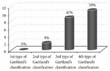
          Purpose. Analysis of early vascular and nerve complications of supracondylar humerus fractures in children. Material and Methods. 220 children hospitalized in the Pediatric Trauma-Orthopedic Department in the years 2004–2014. The group consisted of 143 males and 77 females. Results. Acute neurovascular complications occurred in 16.81% of patients with displaced supracondylar fracture (37 children). Nerve damage was found in 10% of patients with displaced fracture (22 children). The most injured nerve was median nerve; this complication occurred in 15 patients (68.18%). The total nerve function returned after average of 122 days (0–220 days after surgery). Symptoms of vascular injury occurred in 7.7% children with displaced fracture (17 children). Conclusions. (1) In children with supracondylar fracture the most often injured nerve is median nerve. (2) The incidence of vascular and nerve complications positively correlates with the progression of fracture according to Gartland classification.

          Related collections

          Most cited references36

          • Record: found
          • Abstract: found
          • Article: not found

          Supracondylar humeral fractures in children.

          Operative fixation is indicated for most type-II and III supracondylar humeral fractures in order to prevent malunion. Medial comminution is a subtle finding that, if treated nonoperatively, is likely to lead to unacceptable varus malunion. Angiography is not indicated for a pulseless limb, as it delays fracture reduction, which usually corrects the vascular problem. A high index of suspicion is necessary to avoid missing an impending compartment syndrome, especially when there is a concomitant forearm fracture or when there is a median nerve injury, which may mask symptoms of compartment syndrome. Lateral entry pins have been shown, in biomechanical and clinical studies, to be as stable as cross pinning if they are well spaced at the fracture line, and they are not associated with the risk of iatrogenic ulnar nerve injury.
            Bookmark
            • Record: found
            • Abstract: found
            • Article: not found

            Compartment syndrome of the upper extremity.

            Compartment syndrome involves the sustained elevation of interstitial tissue pressures within an osteofascial envelope to nonphysiologic levels. Tissue injury involves a spectrum from reversible to irreversible damage and, therefore, early recognition and treatment is critical for optimal outcomes. This article reviews the nature of upper extremity compartment syndrome; considers the general classification scheme and potential causes; and discusses the pertinent anatomy, pathophysiology, treatment recommendations, and outcomes for this challenging condition. Copyright © 2011. Published by Elsevier Inc.
              Bookmark
              • Record: found
              • Abstract: found
              • Article: not found

              Compartment syndrome of the forearm: a systematic review.

              In this systematic review, we examined the available evidence regarding compartment syndrome of the forearm. Applying our inclusion criteria, we found 12 articles for a total of 84 cases using the MEDLINE (Ovid) database. All were retrospective studies (level IV evidence). In this study, papers were analyzed for causes, diagnosis, treatment, methods of wound closure, functional outcome, and complications. The most common cause of compartment syndrome of the forearm in children was a supracondylar fracture, while in adults the most common cause was a fracture of the distal radius. The diagnostic criterion used was clinical assessment alone in 48%, and in 52%, a combination of measurement of intracompartmental pressure and clinical assessment was used. The intracompartmental pressure was measured using various techniques including a wick catheter, slit catheter, the Whitesides technique, and the Stryker compartment pressure measuring device. Fasciotomy was the preferred method of treatment (73%). In cases reporting wound management, postfasciotomy skin grafting was needed in 61% of the cases, whereas secondary closure was performed in 39% of the cases. Neurological deficit was the most common complication (21%). Copyright © 2011. Published by Elsevier Inc.
                Bookmark

                Author and article information

                Journal
                Biomed Res Int
                Biomed Res Int
                BMRI
                BioMed Research International
                Hindawi
                2314-6133
                2314-6141
                2017
                7 March 2017
                : 2017
                : 2803790
                Affiliations
                Department of Pediatric Traumatology and Orthopedy and Department of Pediatric Surgery and Urology, Silesian Medical University, Katowice, Poland
                Author notes
                *Ryszard Tomaszewski: tomaszewskir@ 123456gmail.com

                Academic Editor: Nikolaos K. Kanakaris

                Author information
                http://orcid.org/0000-0002-1628-0866
                Article
                10.1155/2017/2803790
                5359433
                28367440
                ddeeb496-1290-4578-9c1a-aeed1899dad8
                Copyright © 2017 Ryszard Tomaszewski et al.

                This is an open access article distributed under the Creative Commons Attribution License, which permits unrestricted use, distribution, and reproduction in any medium, provided the original work is properly cited.

                History
                : 19 December 2016
                : 14 February 2017
                Categories
                Clinical Study

                Comments

                Comment on this article