18
views
0
recommends
+1 Recommend
0 collections
    0
    shares
      • Record: found
      • Abstract: found
      • Article: found
      Is Open Access

      microRNA: The Impact on Cancer Stemness and Therapeutic Resistance

      review-article

      Read this article at

      Bookmark
          There is no author summary for this article yet. Authors can add summaries to their articles on ScienceOpen to make them more accessible to a non-specialist audience.

          Abstract

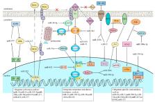
          Cancer ranks as the second leading cause of death worldwide, causing a large social and economic burden. However, most anti-cancer treatments face the problems of tumor recurrence and metastasis. Therefore, finding an effective cure for cancer needs to be solved urgently. Recently, the discovery of cancer stem cells (CSCs) provides a new orientation for cancer research and therapy. CSCs share main characteristics with stem cells and are able to generate an entire tumor. Besides, CSCs usually escape from current anti-cancer therapies, which is partly responsible for tumor recurrence and poor prognosis. microRNAs (miRNAs) belong to small noncoding RNA and regulate gene post-transcriptional expression. The dysregulation of miRNAs leads to plenty of diseases, including cancer. The aberrant miRNA expression in CSCs enhances stemness maintenance. In this review, we summarize the role of miRNAs on CSCs in the eight most common cancers, hoping to bridge the research of miRNAs and CSCs with clinical applications. We found that miRNAs can act as tumor promoter or suppressor. The dysregulation of miRNAs enhances cell stemness and contributes to tumor metastasis and therapeutic resistance via the formation of feedback loops and constitutive activation of carcinogenic signaling pathways. More importantly, some miRNAs may be potential targets for diagnosis, prognosis, and cancer treatments.

          Related collections

          Most cited references175

          • Record: found
          • Abstract: found
          • Article: not found

          Intrinsic resistance of tumorigenic breast cancer cells to chemotherapy.

          Tumorigenic breast cancer cells that express high levels of CD44 and low or undetectable levels of CD24 (CD44(>)/CD24(>/low)) may be resistant to chemotherapy and therefore responsible for cancer relapse. These tumorigenic cancer cells can be isolated from breast cancer biopsies and propagated as mammospheres in vitro. In this study, we aimed to test directly in human breast cancers the effect of conventional chemotherapy or lapatinib (an epidermal growth factor receptor [EGFR]/HER2 pathway inhibitor) on this tumorigenic CD44(>) and CD24(>/low) cell population. Paired breast cancer core biopsies were obtained from patients with primary breast cancer before and after 12 weeks of treatment with neoadjuvant chemotherapy (n = 31) or, for patients with HER2-positive tumors, before and after 6 weeks of treatment with the EGFR/HER2 inhibitor lapatinib (n = 21). Single-cell suspensions established from these biopsies were stained with antibodies against CD24, CD44, and lineage markers and analyzed by flow cytometry. The potential of cells from biopsy samples taken before and after treatment to form mammospheres in culture was compared. All statistical tests were two-sided. Chemotherapy treatment increased the percentage of CD44(>)/CD24(>/low) cells (mean at baseline vs 12 weeks, 4.7%, 95% confidence interval [CI] = 3.5% to 5.9%, vs 13.6%, 95% CI = 10.9% to 16.3%; P )/CD24(>/low) cells (mean at baseline vs 6 weeks, 10.0%, 95% CI = 7.2% to 12.8%, vs 7.5%, 95% CI = 4.1% to 10.9%) and a statistically non-significant decrease in MSFE (mean at baseline vs 6 weeks, 16.1%, 95% CI = 8.7% to 23.5%, vs 10.8%, 95% CI = 4.0% to 17.6%). These studies provide clinical evidence for a subpopulation of chemotherapy-resistant breast cancer-initiating cells. Lapatinib did not lead to an increase in these tumorigenic cells, and, in combination with conventional therapy, specific pathway inhibitors may provide a therapeutic strategy for eliminating these cells to decrease recurrence and improve long-term survival.
            Bookmark
            • Record: found
            • Abstract: found
            • Article: not found

            A protein interaction network for pluripotency of embryonic stem cells.

            Embryonic stem (ES) cells are pluripotent and of therapeutic potential in regenerative medicine. Understanding pluripotency at the molecular level should illuminate fundamental properties of stem cells and the process of cellular reprogramming. Through cell fusion the embryonic cell phenotype can be imposed on somatic cells, a process promoted by the homeodomain protein Nanog, which is central to the maintenance of ES cell pluripotency. Nanog is thought to function in concert with other factors such as Oct4 (ref. 8) and Sox2 (ref. 9) to establish ES cell identity. Here we explore the protein network in which Nanog operates in mouse ES cells. Using affinity purification of Nanog under native conditions followed by mass spectrometry, we have identified physically associated proteins. In an iterative fashion we also identified partners of several Nanog-associated proteins (including Oct4), validated the functional relevance of selected newly identified components and constructed a protein interaction network. The network is highly enriched for nuclear factors that are individually critical for maintenance of the ES cell state and co-regulated on differentiation. The network is linked to multiple co-repressor pathways and is composed of numerous proteins whose encoding genes are putative direct transcriptional targets of its members. This tight protein network seems to function as a cellular module dedicated to pluripotency.
              Bookmark
              • Record: found
              • Abstract: found
              • Article: not found

              A natural antisense transcript regulates Zeb2/Sip1 gene expression during Snail1-induced epithelial-mesenchymal transition.

              Expression of Snail1 in epithelial cells triggers an epithelial-mesenchymal transition (EMT). Here, we demonstrate that the synthesis of Zeb2, a transcriptional repressor of E-cadherin, is up-regulated after Snail1-induced EMT. Snail1 does not affect the synthesis of Zeb2 mRNA, but prevents the processing of a large intron located in its 5'-untranslated region (UTR). This intron contains an internal ribosome entry site (IRES) necessary for the expression of Zeb2. Maintenance of 5'-UTR Zeb2 intron is dependent on the expression of a natural antisense transcript (NAT) that overlaps the 5' splice site in the intron. Ectopic overexpression of this NAT in epithelial cells prevents splicing of the Zeb2 5'-UTR, increases the levels of Zeb2 protein, and consequently down-regulates E-cadherin mRNA and protein. The relevance of these results is demonstrated by the strong association between NAT presence and conservation of the 5'-UTR intron in cells that have undergone EMT or in human tumors with low E-cadherin expression. Therefore, the results presented in this article reveal the existence of a NAT capable of activating Zeb2 expression, explain the mechanism involved in this activation, and demonstrate that this NAT regulates E-cadherin expression.
                Bookmark

                Author and article information

                Journal
                Cells
                Cells
                cells
                Cells
                MDPI
                2073-4409
                18 December 2019
                January 2020
                : 9
                : 1
                : 8
                Affiliations
                Jiangxi Province Key Laboratory of Tumor Pathogenesis and Molecular Pathology, Department of Pathophysiology, Medical College, Nanchang University, 461 Bayi Road, Nanchang 330006, China; jiaoxueqiao1550@ 123456163.com (X.J.); 6302615051@ 123456email.ncu.edu.cn (X.Q.); wulongyuan1201@ 123456163.com (L.W.); libo181818@ 123456126.com (B.L.); 401442719031@ 123456email.ncu.edu.cn (Y.W.); xinyu.kong@ 123456se17.qmul.ac.uk (X.K.)
                Author notes
                [* ]Correspondence: xionglixia@ 123456ncu.edu.cn ; Tel.: +86-791-8636-0556
                [†]

                These authors contributed equally to this work.

                Author information
                https://orcid.org/0000-0002-7193-8077
                https://orcid.org/0000-0001-5366-4976
                https://orcid.org/0000-0002-6751-8715
                https://orcid.org/0000-0002-0616-9718
                https://orcid.org/0000-0003-3769-2496
                Article
                cells-09-00008
                10.3390/cells9010008
                7016867
                31861404
                ddae0d8d-dce3-4c8e-abb7-bf904247e568
                © 2019 by the authors.

                Licensee MDPI, Basel, Switzerland. This article is an open access article distributed under the terms and conditions of the Creative Commons Attribution (CC BY) license ( http://creativecommons.org/licenses/by/4.0/).

                History
                : 16 October 2019
                : 16 December 2019
                Categories
                Review

                microrna,cancer,cancer stem cell,therapeutic resistance,cancer therapy

                Comments

                Comment on this article