15
views
0
recommends
+1 Recommend
0 collections
    0
    shares
      • Record: found
      • Abstract: found
      • Article: found
      Is Open Access

      PD-1, but Not PD-L1, Expressed by Islet-Reactive CD4 + T Cells Suppresses Infiltration of the Pancreas During Type 1 Diabetes

      research-article

      Read this article at

      Bookmark
          There is no author summary for this article yet. Authors can add summaries to their articles on ScienceOpen to make them more accessible to a non-specialist audience.

          Abstract

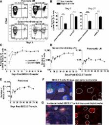
          The inhibitory receptor programmed death-1 (PD-1) constrains type 1 diabetes (T1D) in the nonobese diabetic (NOD) mouse. However, how PD-1 influences diabetogenic CD4 + T cells during natural diabetes is not fully understood. To address this question, we developed a novel model to investigate antigen-specific CD4 + T cells under physiological conditions in vivo. We transferred a low number of naïve CD4 + T cells from the BDC2.5 mouse into prediabetic NOD mice to mimic a physiological precursor frequency and allowed the cells to become primed by endogenous autoantigen. Transferred BDC2.5 T cells became activated, differentiated into T-bet + IFN-γ–producing cells, and infiltrated the pancreas. In this model, loss of PD-1, but not programmed death ligand-1 (PD-L1), on the antigen-specific CD4 + T cell resulted in increased cell numbers in the spleen, pancreas-draining lymph node, and pancreas. PD-1 deficiency also increased expression of the chemokine receptor CXCR3. Lastly, histological data showed that a loss of PD-1 caused BDC2.5 cells to penetrate deep into the islet core, resulting in conversion from peri-insulitis to destructive insulitis. These data support a model by which PD-1 regulates islet-reactive CD4 + T cells in a cell intrinsic manner by suppressing proliferation, inhibiting infiltration of the pancreas, and limiting diabetes.

          Related collections

          Most cited references27

          • Record: found
          • Abstract: found
          • Article: not found

          Foxp3 instability leads to the generation of pathogenic memory T cells in vivo

          Regulatory T (Treg) cells play a central role in maintaining immune homeostasis. However, little is known about the stability of Treg cells in vivo. In this study, we demonstrate that a significant percentage of cells exhibited transient or unstable Foxp3 expression. These exFoxp3+ T cells express an activated-memory T cell phenotype, and produced inflammatory cytokines. Moreover, exFoxp3 cell numbers increased in inflamed tissues under autoimmune conditions. Adoptive transfer of autoreactive exFoxp3 cells led to the rapid-onset of diabetes. Finally, T cell receptor repertoire analyses suggested that exFoxp3 cells develop from both natural and adaptive Treg cells. Thus, the generation of potentially autoreactive effector T cells as a consequence of Foxp3 instability has important implications for understanding autoimmune disease pathogenesis.
            Bookmark
            • Record: found
            • Abstract: found
            • Article: not found

            The Programmed Death-1 (PD-1) Pathway Regulates Autoimmune Diabetes in Nonobese Diabetic (NOD) Mice

            Programmed death-1 (PD-1) receptor, an inhibitory costimulatory molecule found on activated T cells, has been demonstrated to play a role in the regulation of immune responses and peripheral tolerance. We investigated the role of this pathway in the development of autoimmune diabetes. PD-1 or PD-L1 but not PD-L2 blockade rapidly precipitated diabetes in prediabetic female nonobese diabetic (NOD) mice regardless of age (from 1 to 10-wk-old), although it was most pronounced in the older mice. By contrast, cytotoxic T lymphocyte–associated antigen 4 (CTLA-4) blockade induced disease only in neonates. Male NOD mice also developed diabetes after PD-1–PD-L1 pathway blockade, but NOR mice, congenic to NOD but resistant to the development of diabetes, did not. Insulitis scores were significantly higher and frequency of interferon γ–producing GAD-reactive splenocytes was increased after PD-1–PD-L1 pathway blockade compared with controls. Interestingly, PD-L1 but not PD-L2 was found to be expressed on inflamed islets of NOD mice. These data demonstrate a central role for PD-1–PD-L1 interaction in the regulation of induction and progression of autoimmune diabetes in the NOD mouse and provide the rationale to develop new therapies to target this costimulatory pathway in this disease.
              Bookmark
              • Record: found
              • Abstract: found
              • Article: not found

              Blockade of programmed death-1 ligands on dendritic cells enhances T cell activation and cytokine production.

              Programmed death-1 ligand (PD-L)1 and PD-L2 are ligands for programmed death-1 (PD-1), a member of the CD28/CTLA4 family expressed on activated lymphoid cells. PD-1 contains an immunoreceptor tyrosine-based inhibitory motif and mice deficient in PD-1 develop autoimmune disorders suggesting a defect in peripheral tolerance. Human PD-L1 and PD-L2 are expressed on immature dendritic cells (iDC) and mature dendritic cells (mDC), IFN-gamma-treated monocytes, and follicular dendritic cells. Using mAbs, we show that blockade of PD-L2 on dendritic cells results in enhanced T cell proliferation and cytokine production, including that of IFN-gamma and IL-10, while blockade of PD-L1 results in similar, more modest, effects. Blockade of both PD-L1 and PD-L2 showed an additive effect. Both whole mAb and Fab enhanced T cell activation, showing that PD-L1 and PD-L2 function to inhibit T cell activation. Enhancement of T cell activation was most pronounced with weak APC, such as iDCs and IL-10-pretreated mDCs, and less pronounced with strong APC such as mDCs. These data are consistent with the hypothesis that iDC have a balance of stimulatory vs inhibitory molecules that favors inhibition, and indicate that PD-L1 and PD-L2 contribute to the poor stimulatory capacity of iDC. PD-L1 expression differs from PD-L2 in that PD-L1 is expressed on activated T cells, placental trophoblasts, myocardial endothelium, and cortical thymic epithelial cells. In contrast, PD-L2 is expressed on placental endothelium and medullary thymic epithelial cells. PD-L1 is also highly expressed on most carcinomas but minimally expressed on adjacent normal tissue suggesting a role in attenuating antitumor immune responses.
                Bookmark

                Author and article information

                Journal
                Diabetes
                Diabetes
                diabetes
                diabetes
                Diabetes
                Diabetes
                American Diabetes Association
                0012-1797
                1939-327X
                August 2013
                17 July 2013
                : 62
                : 8
                : 2859-2869
                Affiliations
                [1] 1Department of Medicine, Center for Immunology, University of Minnesota, Minneapolis, Minnesota
                [2] 2Department of Microbiology, Center for Immunology, University of Minnesota, Minneapolis, Minnesota
                [3] 3Department of Molecular Immunology, Tokyo Medical and Dental University, Tokyo, Japan
                Author notes
                Corresponding author: Brian T. Fife, bfife@ 123456umn.edu .
                Article
                1475
                10.2337/db12-1475
                3717847
                23545706
                dd58e388-6a24-441b-bb68-1205601581e9
                © 2013 by the American Diabetes Association.

                Readers may use this article as long as the work is properly cited, the use is educational and not for profit, and the work is not altered. See http://creativecommons.org/licenses/by-nc-nd/3.0/ for details.

                History
                : 24 October 2012
                : 21 March 2013
                Page count
                Pages: 11
                Categories
                Original Research
                Immunology and Transplantation

                Endocrinology & Diabetes
                Endocrinology & Diabetes

                Comments

                Comment on this article