11
views
0
recommends
+1 Recommend
0 collections
    0
    shares
      • Record: found
      • Abstract: found
      • Article: found
      Is Open Access

      Mechanobiological Modulation of Cytoskeleton and Calcium Influx in Osteoblastic Cells by Short-Term Focused Acoustic Radiation Force

      research-article
      1 , 2 , 1 , 1 , *
      PLoS ONE
      Public Library of Science

      Read this article at

      Bookmark
          There is no author summary for this article yet. Authors can add summaries to their articles on ScienceOpen to make them more accessible to a non-specialist audience.

          Abstract

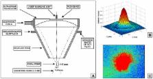
          Mechanotransduction has demonstrated potential for regulating tissue adaptation in vivo and cellular activities in vitro. It is well documented that ultrasound can produce a wide variety of biological effects in biological systems. For example, pulsed ultrasound can be used to noninvasively accelerate the rate of bone fracture healing. Although a wide range of studies has been performed, mechanism for this therapeutic effect on bone healing is currently unknown. To elucidate the mechanism of cellular response to mechanical stimuli induced by pulsed ultrasound radiation, we developed a method to apply focused acoustic radiation force (ARF) (duration, one minute) on osteoblastic MC3T3-E1 cells and observed cellular responses to ARF using a spinning disk confocal microscope. This study demonstrates that the focused ARF induced F-actin cytoskeletal rearrangement in MC3T3-E1 cells. In addition, these cells showed an increase in intracellular calcium concentration following the application of focused ARF. Furthermore, passive bending movement was noted in primary cilium that were treated with focused ARF. Cell viability was not affected. Application of pulsed ultrasound radiation generated only a minimal temperature rise of 0.1°C, and induced a streaming resulting fluid shear stress of 0.186 dyne/cm 2, suggesting that hyperthermia and acoustic streaming might not be the main causes of the observed cell responses. In conclusion, these data provide more insight in the interactions between acoustic mechanical stress and osteoblastic cells. This experimental system could serve as basis for further exploration of the mechanosensing mechanism of osteoblasts triggered by ultrasound.

          Related collections

          Most cited references59

          • Record: found
          • Abstract: found
          • Article: not found

          Cellular mechanotransduction: putting all the pieces together again.

          Analysis of cellular mechanotransduction, the mechanism by which cells convert mechanical signals into biochemical responses, has focused on identification of critical mechanosensitive molecules and cellular components. Stretch-activated ion channels, caveolae, integrins, cadherins, growth factor receptors, myosin motors, cytoskeletal filaments, nuclei, extracellular matrix, and numerous other structures and signaling molecules have all been shown to contribute to the mechanotransduction response. However, little is known about how these different molecules function within the structural context of living cells, tissues, and organs to produce the orchestrated cellular behaviors required for mechanosensation, embryogenesis, and physiological control. Recent work from a wide range of fields reveals that organ, tissue, and cell anatomy are as important for mechanotransduction as individual mechanosensitive proteins and that our bodies use structural hierarchies (systems within systems) composed of interconnected networks that span from the macroscale to the nanoscale in order to focus stresses on specific mechanotransducer molecules. The presence of isometric tension (prestress) at all levels of these multiscale networks ensures that various molecular scale mechanochemical transduction mechanisms proceed simultaneously and produce a concerted response. Future research in this area will therefore require analysis, understanding, and modeling of tensionally integrated (tensegrity) systems of mechanochemical control.
            Bookmark
            • Record: found
            • Abstract: found
            • Article: not found

            A model for the excitation of osteocytes by mechanical loading-induced bone fluid shear stresses.

            A new experimentally testable hypothesis is advanced for the mechanosensory transduction mechanism by which communicating osteocytes sense the very small in vivo strains in the calcified matrix components of bone. We propose that the osteocytes, although not responsive to substantial fluid pressures, can be stimulated by relatively small fluid shear stresses acting on the membranes of their osteocytic processes. Biot's porous media theory is used to relate the combined axial and bending loads applied to a whole bone to the flow past the osteocytic processes in their canaliculi. In this theory, the bone pores of interest are the proteoglycan filled fluid annuli that surround the osteocytic processes in the canaliculi. We show that previously predicted fluid pore pressure relaxation times were a hundred-fold too short for the lacunar-canalicular porosity because they neglected the fluid drag associated with proteoglycan matrix on the surface membrane of the osteocyte and its cell processes. The recent theory developed in Tsay and Weinbaum [J. Fluid Mech. 226, 125-148 (1991)] for flow through cross-linked fiber filled channels is used to model the flow through this proteoglycan matrix. The predicted pore relaxation time, 1-2 s, closely corresponds to the times measured by Salzstein and Pollack [J. Biomechanics 20, 271-280 (1987)]. Furthermore, using this model, the magnitude of the predicted fluid induced shear stresses, 8-30 dyn cm-2, is shown to be similar to the fluid shear stresses measured in osteoblasts and other cells in which an intracellular Ca2+ shear stress response had been observed. This model is also used, in conjunction with anatomical data and the pore fluid pressure relaxation time data, to show that the spacing between the fibers is approximately 7 nm. The result is consistent with the notion that the canalicular pore space is filled with glycosaminoglycans that are ordered by albumin according to the model of Michel [J. Physiol. 404, 1-29 (1988)]. The new hypothesis is also shown to be consistent with the experiments of McLeod et al. [J. Biomechanics (submitted)] which suggest that high-frequency low-amplitude postural strains can maintain and even increase bone mass.
              Bookmark
              • Record: found
              • Abstract: found
              • Article: not found

              Mechanical bioeffects of ultrasound.

              Ultrasound is used widely in medicine as both a diagnostic and therapeutic tool. Through both thermal and nonthermal mechanisms, ultrasound can produce a variety of biological effects in tissues in vitro and in vivo. This chapter provides an overview of the fundamentals of key nonthermal mechanisms for the interaction of ultrasound with biological tissues. Several categories of mechanical bioeffects of ultrasound are then reviewed to provide insight on the range of ultrasound bioeffects in vivo, the relevance of these effects to diagnostic imaging, and the potential application of mechanical bioeffects to the design of new therapeutic applications of ultrasound in medicine.
                Bookmark

                Author and article information

                Contributors
                Role: Editor
                Journal
                PLoS One
                PLoS ONE
                plos
                plosone
                PLoS ONE
                Public Library of Science (San Francisco, USA )
                1932-6203
                2012
                6 June 2012
                : 7
                : 6
                : e38343
                Affiliations
                [1 ]Department of Biomedical Engineering, Stony Brook University, Stony Brook, New York, United States of America
                [2 ]The Key Laboratory of Aerospace Medicine, Chinese Ministry of Education, Xi’an, ShaanXi, People's Republic of China
                University of Texas Southwestern Medical Center, United States of America
                Author notes

                Conceived and designed the experiments: SZ JQC YXQ. Performed the experiments: SZ JQC YXQ. Analyzed the data: SZ JQC YXQ. Contributed reagents/materials/analysis tools: SZ JQC YXQ. Wrote the paper: SZ JQC YXQ.

                Article
                PONE-D-11-26111
                10.1371/journal.pone.0038343
                3368843
                22701628
                dbdc6fa6-4bd0-4ff1-a691-89622ccc139a
                Zhang et al. This is an open-access article distributed under the terms of the Creative Commons Attribution License, which permits unrestricted use, distribution, and reproduction in any medium, provided the original author and source are credited.
                History
                : 20 December 2011
                : 3 May 2012
                Page count
                Pages: 11
                Categories
                Research Article
                Biology
                Anatomy and Physiology
                Musculoskeletal System
                Biomechanics
                Bone and Joint Mechanics
                Cell Mechanics
                Bone
                Physiological Processes
                Biomineralization
                Homeostasis
                Biophysics
                Cell Motility
                Ciliary Movement
                Biomechanics
                Biophysics Simulations
                Biotechnology
                Bioengineering
                Biomedical Engineering
                Tissue Engineering

                Uncategorized
                Uncategorized

                Comments

                Comment on this article