3
views
0
recommends
+1 Recommend
0 collections
    0
    shares
      • Record: found
      • Abstract: found
      • Article: found
      Is Open Access

      Effect of APOE ε4 genotype on amyloid-β and tau accumulation in Alzheimer’s disease

      research-article

      Read this article at

      Bookmark
          There is no author summary for this article yet. Authors can add summaries to their articles on ScienceOpen to make them more accessible to a non-specialist audience.

          Abstract

          Background

          To assess the effects of apolipoprotein E (ApoE) ε4 genotype on amyloid-β (Aβ) and tau burden and their longitudinal changes in Alzheimer’s disease (AD) spectrum.

          Methods

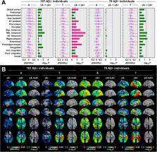
          Among 272 individuals who underwent PET scans ( 18F-florbetaben for Aβ and 18F-flortaucipir for tau) and ApoE genotyping, 187 individuals completed 2-year follow-up PET scans. After correcting for the partial volume effect, we compared the standardized uptake value ratio (SUVR) for Aβ and tau burden between the ε4+ and ε4− groups. By using a linear mixed-effect model, we measured changes in SUVR in the ApoE ε4+ and ε4− groups.

          Results

          The ε4+ group showed greater baseline Aβ burden in the diffuse cortical regions and greater tau burden in the lateral, and medial temporal, cingulate, and insula cortices. Tau accumulation rate was higher in the parietal, occipital, lateral, and medial temporal cortices in the ε4+ group. In Aβ+ individuals, baseline tau burden was greater in the medial temporal cortex, while Aβ burden was conversely greater in the ε4− group. Tau accumulation rate was higher in the ε4+ group in a small region in the lateral temporal cortex. The effect of ApoE ε4 on enhanced tau accumulation persisted even after adjusting for the global cortical Aβ burden.

          Conclusions

          Progressive tau accumulation may be more prominent in ε4 carriers, particularly in the medial and lateral temporal cortices. ApoE ε4 allele has differential effects on the Aβ burden depending on the existing amyloidosis and may enhance vulnerability to progressive tau accumulation in the AD spectrum independent of Aβ.

          Supplementary information

          Supplementary information accompanies this paper at 10.1186/s13195-020-00710-6.

          Related collections

          Most cited references50

          • Record: found
          • Abstract: not found
          • Article: not found

          Controlling the False Discovery Rate: A Practical and Powerful Approach to Multiple Testing

            Bookmark
            • Record: found
            • Abstract: found
            • Article: not found

            An automated labeling system for subdividing the human cerebral cortex on MRI scans into gyral based regions of interest.

            In this study, we have assessed the validity and reliability of an automated labeling system that we have developed for subdividing the human cerebral cortex on magnetic resonance images into gyral based regions of interest (ROIs). Using a dataset of 40 MRI scans we manually identified 34 cortical ROIs in each of the individual hemispheres. This information was then encoded in the form of an atlas that was utilized to automatically label ROIs. To examine the validity, as well as the intra- and inter-rater reliability of the automated system, we used both intraclass correlation coefficients (ICC), and a new method known as mean distance maps, to assess the degree of mismatch between the manual and the automated sets of ROIs. When compared with the manual ROIs, the automated ROIs were highly accurate, with an average ICC of 0.835 across all of the ROIs, and a mean distance error of less than 1 mm. Intra- and inter-rater comparisons yielded little to no difference between the sets of ROIs. These findings suggest that the automated method we have developed for subdividing the human cerebral cortex into standard gyral-based neuroanatomical regions is both anatomically valid and reliable. This method may be useful for both morphometric and functional studies of the cerebral cortex as well as for clinical investigations aimed at tracking the evolution of disease-induced changes over time, including clinical trials in which MRI-based measures are used to examine response to treatment.
              Bookmark
              • Record: found
              • Abstract: found
              • Article: not found

              Clinical diagnosis of Alzheimer's disease: Report of the NINCDS-ADRDA Work Group* under the auspices of Department of Health and Human Services Task Force on Alzheimer's Disease

              Neurology, 34(7), 939-939
                Bookmark

                Author and article information

                Contributors
                lyoochel@yuhs.ac
                Journal
                Alzheimers Res Ther
                Alzheimers Res Ther
                Alzheimer's Research & Therapy
                BioMed Central (London )
                1758-9193
                31 October 2020
                31 October 2020
                2020
                : 12
                : 140
                Affiliations
                [1 ]GRID grid.15444.30, ISNI 0000 0004 0470 5454, Department of Neurology, Gangnam Severance Hospital, , Yonsei University College of Medicine, ; 20 Eonjuro 63-gil, Gangnam-gu, Seoul, South Korea
                [2 ]GRID grid.15444.30, ISNI 0000 0004 0470 5454, Biostatistics Collaboration Unit, Gangnam Severance Hospital, , Yonsei University College of Medicine, ; Seoul, South Korea
                [3 ]GRID grid.15444.30, ISNI 0000 0004 0470 5454, Department of Nuclear Medicine, Gangnam Severance Hospital, , Yonsei University College of Medicine, ; 20 Eonjuro 63-gil, Gangnam-gu, Seoul, South Korea
                Author information
                http://orcid.org/0000-0003-2231-672X
                Article
                710
                10.1186/s13195-020-00710-6
                7603688
                33129364
                dbbb24ba-2749-4635-a699-bf8e6abc771f
                © The Author(s) 2020

                Open AccessThis article is licensed under a Creative Commons Attribution 4.0 International License, which permits use, sharing, adaptation, distribution and reproduction in any medium or format, as long as you give appropriate credit to the original author(s) and the source, provide a link to the Creative Commons licence, and indicate if changes were made. The images or other third party material in this article are included in the article's Creative Commons licence, unless indicated otherwise in a credit line to the material. If material is not included in the article's Creative Commons licence and your intended use is not permitted by statutory regulation or exceeds the permitted use, you will need to obtain permission directly from the copyright holder. To view a copy of this licence, visit http://creativecommons.org/licenses/by/4.0/. The Creative Commons Public Domain Dedication waiver ( http://creativecommons.org/publicdomain/zero/1.0/) applies to the data made available in this article, unless otherwise stated in a credit line to the data.

                History
                : 7 August 2020
                : 20 October 2020
                Funding
                Funded by: 2020 Research Grant of Gangnam Severance Hospital Research Committee
                Funded by: Yonsei University College of Medicine (KR)
                Award ID: 6-2018-0068
                Award Recipient :
                Funded by: FundRef http://dx.doi.org/10.13039/501100003725, National Research Foundation of Korea;
                Award ID: NRF2020R1F1A1076154
                Award ID: NRF2018R1D1A1B07049386
                Award Recipient :
                Funded by: FundRef http://dx.doi.org/10.13039/501100003710, Korea Health Industry Development Institute;
                Award ID: HI18C1159
                Award Recipient :
                Categories
                Research
                Custom metadata
                © The Author(s) 2020

                Neurology
                alzheimer disease,amyloid-β,apoe,positron emission tomography,tau
                Neurology
                alzheimer disease, amyloid-β, apoe, positron emission tomography, tau

                Comments

                Comment on this article