3
views
0
recommends
+1 Recommend
0 collections
    0
    shares
      • Record: found
      • Abstract: found
      • Article: found
      Is Open Access

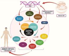
      Pseudogenes and Liquid Phase Separation in Epigenetic Expression

      review-article

      Read this article at

      Bookmark
          There is no author summary for this article yet. Authors can add summaries to their articles on ScienceOpen to make them more accessible to a non-specialist audience.

          Abstract

          Pseudogenes have been considered as non-functional genes. However, peptides and long non-coding RNAs produced by pseudogenes are expressed in different tumors. Moreover, the dysregulation of pseudogenes is associated with cancer, and their expressions are higher in tumors compared to normal tissues. Recent studies show that pseudogenes can influence the liquid phase condensates formation. Liquid phase separation involves regulating different epigenetic stages, including transcription, chromatin organization, 3D DNA structure, splicing, and post-transcription modifications like m 6A. Several membrane-less organelles, formed through the liquid phase separate, are also involved in the epigenetic regulation, and their defects are associated with cancer development. However, the association between pseudogenes and liquid phase separation remains unrevealed. The current study sought to investigate the relationship between pseudogenes and liquid phase separation in cancer development, as well as their therapeutic implications.

          Related collections

          Most cited references98

          • Record: found
          • Abstract: found
          • Article: not found

          Coactivator condensation at super-enhancers links phase separation and gene control

          Super-enhancers (SEs) are clusters of enhancers that cooperatively assemble a high density of transcriptional apparatus to drive robust expression of genes with prominent roles in cell identity. Here, we demonstrate that the SE-enriched transcriptional coactivators BRD4 and MED1 form nuclear puncta at SEs that exhibit properties of liquid-like condensates and are disrupted by chemicals that perturb condensates. The intrinsically disordered regions (IDRs) of BRD4 and MED1 can form phase-separated droplets and MED1-IDR droplets can compartmentalize and concentrate transcription apparatus from nuclear extracts. These results support the idea that coactivators form phase-separated condensates at SEs that compartmentalize and concentrate the transcription apparatus, suggest a role for coactivator IDRs in this process, and offer insights into mechanisms involved in control of key cell identity genes.
            Bookmark
            • Record: found
            • Abstract: found
            • Article: not found

            Organization of Chromatin by Intrinsic and Regulated Phase Separation

            Eukaryotic chromatin is highly condensed yet dynamically accessible to regulation, and organized into subdomains. We demonstrate that reconstituted chromatin undergoes histone tail-driven liquid-liquid phase separation (LLPS) in physiologic salt and when microinjected into cell nuclei, producing dense and dynamic droplets. Linker histone H1 and internucleosome linker lengths shared across eukaryotes promote phase separation of chromatin, tune droplet properties, and coordinate to form condensates of consistent density, in manners that parallel chromatin behaviors in cells. Histone acetylation by p300 antagonizes chromatin phase separation, dissolving droplets in vitro and decreasing droplet formation in nuclei. In the presence of multi-bromodomain proteins, such as BRD4, highly acetylated chromatin forms a new phase separated state, with droplets of distinct physical properties, which can be immiscible with unmodified chromatin droplets, mimicking nuclear chromatin subdomains. Our data suggest a framework, based on intrinsic phase separation of the chromatin polymer, for understanding organization and regulation of eukaryotic genomes.
              Bookmark
              • Record: found
              • Abstract: found
              • Article: not found

              The nucleolus as a multiphase liquid condensate

              The nucleolus is the most prominent nuclear body and serves a fundamentally important biological role as a site of ribonucleoprotein particle assembly, primarily dedicated to ribosome biogenesis. Despite being one of the first intracellular structures visualized historically, the biophysical rules governing its assembly and function are only starting to become clear. Recent studies have provided increasing support for the concept that the nucleolus represents a multilayered biomolecular condensate, whose formation by liquid-liquid phase separation (LLPS) facilitates the initial steps of ribosome biogenesis and other functions. Here, we review these biophysical insights in the context of the molecular and cell biology of the nucleolus. We discuss how nucleolar function is linked to its organization as a multiphase condensate and how dysregulation of this organization could provide insights into still poorly understood aspects of nucleolus-associated diseases, including cancer, ribosomopathies and neurodegeneration as well as ageing. We suggest that the LLPS model provides the starting point for a unifying quantitative framework for the assembly, structural maintenance and function of the nucleolus, with implications for gene regulation and ribonucleoprotein particle assembly throughout the nucleus. The LLPS concept is also likely useful in designing new therapeutic strategies to target nucleolar dysfunction.
                Bookmark

                Author and article information

                Contributors
                Journal
                Front Oncol
                Front Oncol
                Front. Oncol.
                Frontiers in Oncology
                Frontiers Media S.A.
                2234-943X
                08 July 2022
                2022
                : 12
                : 912282
                Affiliations
                [1] 1 Laboratory of Cell Signal Transduction, Department of Biochemistry and Molecular Biology, School of Basic Medical Sciences, Henan University , Kaifeng, China
                [2] 2 School of Life Sciences, Henan University , Kaifeng, China
                [3] 3 Department of Basic Sciences Research, Shaukat Khanum Memorial Cancer Hospital and Research Centre (SKMCH&RC) , Lahore, Pakistan
                [4] 4 Department of Medical Laboratory Technology, The University of Haripur , Haripur, Pakistan
                Author notes

                Edited by: Kevin J Ni, St George Hospital Cancer Care Centre, Australia

                Reviewed by: Pradeep Reddy Cingaram, University of Miami, United States

                *Correspondence: Wenqiang Wei, weiwq@ 123456henu.edu.cn ; Weijuan Zhang, zwjuan1965@ 123456henu.edu.cn ; Shaoping Ji, shaopingji@ 123456henu.edu.cn

                †These authors have contributed equally to this work and share first authorship

                This article was submitted to Molecular and Cellular Oncology, a section of the journal Frontiers in Oncology

                Article
                10.3389/fonc.2022.912282
                9305658
                35875144
                dbb39b6a-fc3a-4df3-b141-f123b1e5a3b0
                Copyright © 2022 Nsengimana, Khan, Awan, Wang, Fang, Wei, Zhang and Ji

                This is an open-access article distributed under the terms of the Creative Commons Attribution License (CC BY). The use, distribution or reproduction in other forums is permitted, provided the original author(s) and the copyright owner(s) are credited and that the original publication in this journal is cited, in accordance with accepted academic practice. No use, distribution or reproduction is permitted which does not comply with these terms.

                History
                : 04 April 2022
                : 13 June 2022
                Page count
                Figures: 2, Tables: 1, Equations: 0, References: 99, Pages: 11, Words: 4927
                Funding
                Funded by: National Natural Science Foundation of China , doi 10.13039/501100001809;
                Categories
                Oncology
                Mini Review

                Oncology & Radiotherapy
                liquid phase separation,pseudogenes,rna modification,epigenetic,cancer
                Oncology & Radiotherapy
                liquid phase separation, pseudogenes, rna modification, epigenetic, cancer

                Comments

                Comment on this article