6
views
0
recommends
+1 Recommend
0 collections
    0
    shares
      • Record: found
      • Abstract: found
      • Article: found
      Is Open Access

      Integrative analysis of multi-platform reverse-phase protein array data for the pharmacodynamic assessment of response to targeted therapies

      research-article

      Read this article at

      Bookmark
          There is no author summary for this article yet. Authors can add summaries to their articles on ScienceOpen to make them more accessible to a non-specialist audience.

          Abstract

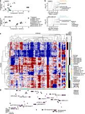
          Reverse-phase protein array (RPPA) technology uses panels of high-specificity antibodies to measure proteins and protein post-translational modifications in cells and tissues. The approach offers sensitive and precise quantification of large numbers of samples and has thus found applications in the analysis of clinical and pre-clinical samples. For effective integration into drug development and clinical practice, robust assays with consistent results are essential. Leveraging a collaborative RPPA model, we set out to assess the variability between three different RPPA platforms using distinct instrument set-ups and workflows. Employing multiple RPPA-based approaches operated across distinct laboratories, we characterised a range of human breast cancer cells and their protein-level responses to two clinically relevant cancer drugs. We integrated multi-platform RPPA data and used unsupervised learning to identify protein expression and phosphorylation signatures that were not dependent on RPPA platform and analysis workflow. Our findings indicate that proteomic analyses of cancer cell lines using different RPPA platforms can identify concordant profiles of response to pharmacological inhibition, including when using different antibodies to measure the same target antigens. These results highlight the robustness and the reproducibility of RPPA technology and its capacity to identify protein markers of disease or response to therapy.

          Related collections

          Most cited references59

          • Record: found
          • Abstract: found
          • Article: not found

          The Perseus computational platform for comprehensive analysis of (prote)omics data.

          A main bottleneck in proteomics is the downstream biological analysis of highly multivariate quantitative protein abundance data generated using mass-spectrometry-based analysis. We developed the Perseus software platform (http://www.perseus-framework.org) to support biological and biomedical researchers in interpreting protein quantification, interaction and post-translational modification data. Perseus contains a comprehensive portfolio of statistical tools for high-dimensional omics data analysis covering normalization, pattern recognition, time-series analysis, cross-omics comparisons and multiple-hypothesis testing. A machine learning module supports the classification and validation of patient groups for diagnosis and prognosis, and it also detects predictive protein signatures. Central to Perseus is a user-friendly, interactive workflow environment that provides complete documentation of computational methods used in a publication. All activities in Perseus are realized as plugins, and users can extend the software by programming their own, which can be shared through a plugin store. We anticipate that Perseus's arsenal of algorithms and its intuitive usability will empower interdisciplinary analysis of complex large data sets.
            Bookmark
            • Record: found
            • Abstract: found
            • Article: not found
            Is Open Access

            Comprehensive molecular portraits of human breast tumors

            Summary We analyzed primary breast cancers by genomic DNA copy number arrays, DNA methylation, exome sequencing, mRNA arrays, microRNA sequencing and reverse phase protein arrays. Our ability to integrate information across platforms provided key insights into previously-defined gene expression subtypes and demonstrated the existence of four main breast cancer classes when combining data from five platforms, each of which shows significant molecular heterogeneity. Somatic mutations in only three genes (TP53, PIK3CA and GATA3) occurred at > 10% incidence across all breast cancers; however, there were numerous subtype-associated and novel gene mutations including the enrichment of specific mutations in GATA3, PIK3CA and MAP3K1 with the Luminal A subtype. We identified two novel protein expression-defined subgroups, possibly contributed by stromal/microenvironmental elements, and integrated analyses identified specific signaling pathways dominant in each molecular subtype including a HER2/p-HER2/HER1/p-HER1 signature within the HER2-Enriched expression subtype. Comparison of Basal-like breast tumors with high-grade Serous Ovarian tumors showed many molecular commonalities, suggesting a related etiology and similar therapeutic opportunities. The biologic finding of the four main breast cancer subtypes caused by different subsets of genetic and epigenetic abnormalities raises the hypothesis that much of the clinically observable plasticity and heterogeneity occurs within, and not across, these major biologic subtypes of breast cancer.
              Bookmark
              • Record: found
              • Abstract: found
              • Article: not found

              Gene expression patterns of breast carcinomas distinguish tumor subclasses with clinical implications.

              The purpose of this study was to classify breast carcinomas based on variations in gene expression patterns derived from cDNA microarrays and to correlate tumor characteristics to clinical outcome. A total of 85 cDNA microarray experiments representing 78 cancers, three fibroadenomas, and four normal breast tissues were analyzed by hierarchical clustering. As reported previously, the cancers could be classified into a basal epithelial-like group, an ERBB2-overexpressing group and a normal breast-like group based on variations in gene expression. A novel finding was that the previously characterized luminal epithelial/estrogen receptor-positive group could be divided into at least two subgroups, each with a distinctive expression profile. These subtypes proved to be reasonably robust by clustering using two different gene sets: first, a set of 456 cDNA clones previously selected to reflect intrinsic properties of the tumors and, second, a gene set that highly correlated with patient outcome. Survival analyses on a subcohort of patients with locally advanced breast cancer uniformly treated in a prospective study showed significantly different outcomes for the patients belonging to the various groups, including a poor prognosis for the basal-like subtype and a significant difference in outcome for the two estrogen receptor-positive groups.
                Bookmark

                Author and article information

                Contributors
                adam.byron@igmm.ed.ac.uk
                leanne.de-koning@curie.fr
                Journal
                Sci Rep
                Sci Rep
                Scientific Reports
                Nature Publishing Group UK (London )
                2045-2322
                15 December 2020
                15 December 2020
                2020
                : 10
                : 21985
                Affiliations
                [1 ]GRID grid.4305.2, ISNI 0000 0004 1936 7988, Cancer Research UK Edinburgh Centre, Institute of Genetics and Molecular Medicine, , University of Edinburgh, ; Crewe Road South, Edinburgh, EH4 2XR UK
                [2 ]GRID grid.7497.d, ISNI 0000 0004 0492 0584, Division of Molecular Genome Analysis, , German Cancer Research Center (DKFZ), ; Heidelberg, Germany
                [3 ]GRID grid.440907.e, ISNI 0000 0004 1784 3645, Department of Translational Research, Institut Curie, , PSL Research University, ; 26 rue d’Ulm, 75005 Paris, France
                [4 ]GRID grid.440907.e, ISNI 0000 0004 1784 3645, U900 INSERM, Institut Curie, , PSL Research University, ; Paris, France
                [5 ]GRID grid.476393.c, ISNI 0000 0004 4904 8590, Present Address: Pfizer Pharma GmbH, ; Berlin, Germany
                [6 ]Present Address: Sederma, Le Perray-en-Yvelines, France
                [7 ]GRID grid.410368.8, ISNI 0000 0001 2191 9284, Present Address: U1236 INSERM, Faculté de Médecine, , Université de Rennes 1, ; Rennes, France
                [8 ]Present Address: NanoString Technologies, Inc., Seattle, WA USA
                Author information
                http://orcid.org/0000-0002-5939-9883
                http://orcid.org/0000-0002-8817-3672
                http://orcid.org/0000-0001-5541-9747
                http://orcid.org/0000-0003-1826-4584
                http://orcid.org/0000-0002-8072-3633
                Article
                77335
                10.1038/s41598-020-77335-0
                7738515
                33319783
                dba83327-7a21-4aac-b058-c17f4b760658
                © The Author(s) 2020

                Open Access This article is licensed under a Creative Commons Attribution 4.0 International License, which permits use, sharing, adaptation, distribution and reproduction in any medium or format, as long as you give appropriate credit to the original author(s) and the source, provide a link to the Creative Commons licence, and indicate if changes were made. The images or other third party material in this article are included in the article's Creative Commons licence, unless indicated otherwise in a credit line to the material. If material is not included in the article's Creative Commons licence and your intended use is not permitted by statutory regulation or exceeds the permitted use, you will need to obtain permission directly from the copyright holder. To view a copy of this licence, visit http://creativecommons.org/licenses/by/4.0/.

                History
                : 18 September 2019
                : 11 March 2020
                Funding
                Funded by: FundRef https://doi.org/10.13039/501100000289, Cancer Research UK;
                Funded by: Cancéropôle Île-de-France
                Categories
                Article
                Custom metadata
                © The Author(s) 2020

                Uncategorized
                biomarkers,data integration,cancer,tumour biomarkers,biological techniques,proteomic analysis

                Comments

                Comment on this article