3
views
0
recommends
+1 Recommend
0 collections
    0
    shares
      • Record: found
      • Abstract: found
      • Article: found
      Is Open Access

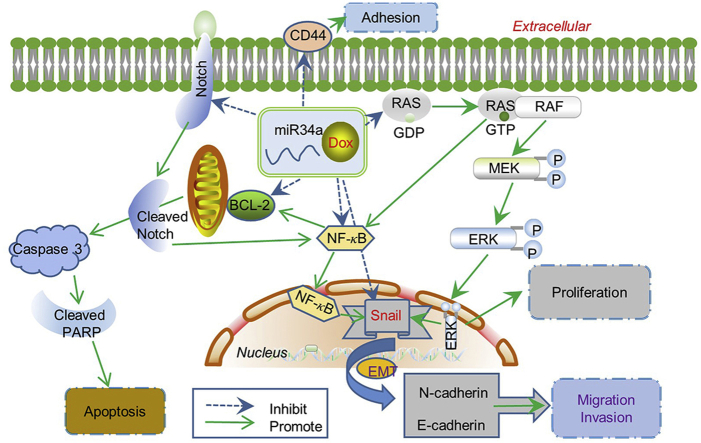
      Combination therapy with miR34a and doxorubicin synergistically inhibits Dox-resistant breast cancer progression via down-regulation of Snail through suppressing Notch/NF- κB and RAS/RAF/MEK/ERK signaling pathway

      research-article

      Read this article at

      Bookmark
          There is no author summary for this article yet. Authors can add summaries to their articles on ScienceOpen to make them more accessible to a non-specialist audience.

          Abstract

          Resistance to breast cancer (BCa) chemotherapy severely hampers the patient's prognosis. MicroRNAs provide a potential therapeutic prospect for BCa. In this study, the reversal function of microRNA34a (miR34a) on doxorubicin (Dox) resistance of BCa and the possible mechanism was investigated. We found that the relative level of miR34a was significantly decreased in Dox-resistant breast cancer cell MCF-7 (MCF-7/A) compared with Dox-sensitive MCF-7 cells. Transfection with miR34a significantly suppressed the invasion, migration, adhesion of MCF-7/A cells without inhibiting their growth obviously. The combination of miR34a and Dox could significantly inhibit the proliferation, migration, invasion and induce the apoptosis of MCF-7/A cells. The synergistic effect of this combination on resistant MCF-7/A cells has no obvious relation with the expressions of classical drug-resistant proteins P-GP, MRP and GST- π, while closely related with the down-regulation on TOP2A and BCRP. Moreover, we found both protein and mRNA expression of Snail were significantly up-regulated in MCF-7/A cells in comparison with MCF-7 cells. Transfection with small interfering RNA (siRNA) of Snail could inhibit the invasion, migration and adhesion of drug-resistant MCF-7/A cells, while high-expression of Snail could remarkably promote the invasion, migration and adhesion of MCF-7 cells, which might be related with regulation of N-cadherin and E-cadherin. Transfection with miR34a in MCF-7/A cells induced a decrease of Snail expression. The potential binding sites of miR34a with 3′ UTR of Snail were predicted by miRDB target prediction software, which was confirmed by luciferase reporter gene method. Results showed that the relative activity of luciferase was reduced in MCF-7/A cells after co-transfection of miR34a and wild type (wt)-Snail, while did not change by co-transfection with miR34a and 3′ UTR mutant type (mut) Snail. Combination of miR34a and Dox induced a stronger decrease of Snail in MCF-7/A cells in comparison to miR34a or Dox treatment alone. What’ more, for the first time, we also found miR34a combined with Dox could obviously inhibit the expression of Snail through suppressing Notch/NF- κB and RAS/RAF/MEK/ERK pathway in MCF-7/A cells. In vivo study indicated that combination of miR34a and Dox significantly slowed down tumor growth in MCF-7/A nude mouse xenograft model compared with Dox alone, which was manifested by the down-regulation of Snail and pro-apoptosis effect in tumor xenografts. These results together underline the relevance of miR34a-driven regulation of Snail in drug resistance and co-administration of miR34a and Dox may produce an effective therapy outcome in the future in clinic.

          Graphical abstract

          Combination of miR34a and doxorubicin synergistically inhibits the proliferation, migration and invasion, and promotes apoptosis of MCF-7/A cells, which may be associated with down-regulation Snail through Notch/NF- κB and RAS/RAF/MEK/ERK pathways.

          Related collections

          Most cited references56

          • Record: found
          • Abstract: found
          • Article: not found

          Molecular mechanisms of epithelial-mesenchymal transition.

          The transdifferentiation of epithelial cells into motile mesenchymal cells, a process known as epithelial-mesenchymal transition (EMT), is integral in development, wound healing and stem cell behaviour, and contributes pathologically to fibrosis and cancer progression. This switch in cell differentiation and behaviour is mediated by key transcription factors, including SNAIL, zinc-finger E-box-binding (ZEB) and basic helix-loop-helix transcription factors, the functions of which are finely regulated at the transcriptional, translational and post-translational levels. The reprogramming of gene expression during EMT, as well as non-transcriptional changes, are initiated and controlled by signalling pathways that respond to extracellular cues. Among these, transforming growth factor-β (TGFβ) family signalling has a predominant role; however, the convergence of signalling pathways is essential for EMT.
            Bookmark
            • Record: found
            • Abstract: found
            • Article: not found

            EMT Transition States during Tumor Progression and Metastasis

            Epithelial-mesenchymal transition (EMT) is a process in which epithelial cells acquire mesenchymal features. In cancer, EMT is associated with tumor initiation, invasion, metastasis, and resistance to therapy. Recently, it has been demonstrated that EMT is not a binary process, but occurs through distinct cellular states. Here, we review the recent studies that demonstrate the existence of these different EMT states in cancer and the mechanisms regulating their functions. We discuss the different functional characteristics, such as proliferation, propagation, plasticity, invasion, and metastasis associated with the distinct EMT states. We summarize the role of the transcriptional and epigenetic landscapes, gene regulatory network and their surrounding niche in controlling the transition through the different EMT states.
              Bookmark
              • Record: found
              • Abstract: found
              • Article: not found

              EMT, MET, Plasticity, and Tumor Metastasis.

              Cancer cell identity and plasticity are required in transition states, such as epithelial-mesenchymal transition (EMT) and mesenchymal-epithelial transition (MET), in primary tumor initiation, progression, and metastasis. The functional roles of EMT, MET, and the partial state (referred to as pEMT) may vary based on the type of tumor, the state of dissemination, and the degree of metastatic colonization. Herein, we review EMT, MET, pEMT, and plasticity in the context of tumor metastasis.
                Bookmark

                Author and article information

                Contributors
                Journal
                Acta Pharm Sin B
                Acta Pharm Sin B
                Acta Pharmaceutica Sinica. B
                Elsevier
                2211-3835
                2211-3843
                08 June 2021
                September 2021
                08 June 2021
                : 11
                : 9
                : 2819-2834
                Affiliations
                [1]Department of Pharmacology, Key Laboratory of Chemical Biology (Ministry of Education), Drug Screening Unit Platform, School of Pharmaceutical Sciences, Shandong University, Jinan 250012, China
                Author notes
                []Corresponding author. Tel./fax: +86 531 88382490. guoxl@ 123456sdu.edu.cn
                Article
                S2211-3835(21)00215-X
                10.1016/j.apsb.2021.06.003
                8463267
                34589399
                db8110b8-27e1-4114-8853-99388f401049
                © 2021 Chinese Pharmaceutical Association and Institute of Materia Medica, Chinese Academy of Medical Sciences. Production and hosting by Elsevier B.V.

                This is an open access article under the CC BY-NC-ND license (http://creativecommons.org/licenses/by-nc-nd/4.0/).

                History
                : 29 January 2021
                : 24 April 2021
                : 19 May 2021
                Categories
                Original Article

                breast cancer,mir34a,dox,drug resistance,snail,notch/nf-κb,ras/raf/mek/erk,therapy

                Comments

                Comment on this article