12
views
0
recommends
+1 Recommend
0 collections
    0
    shares
      • Record: found
      • Abstract: found
      • Article: found
      Is Open Access

      Target identification of natural medicine with chemical proteomics approach: probe synthesis, target fishing and protein identification

      review-article

      Read this article at

      Bookmark
          There is no author summary for this article yet. Authors can add summaries to their articles on ScienceOpen to make them more accessible to a non-specialist audience.

          Abstract

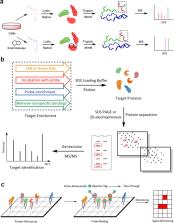
          Natural products are an important source of new drugs for the treatment of various diseases. However, developing natural product-based new medicines through random moiety modification is a lengthy and costly process, due in part to the difficulties associated with comprehensively understanding the mechanism of action and the side effects. Identifying the protein targets of natural products is an effective strategy, but most medicines interact with multiple protein targets, which complicate this process. In recent years, an increasing number of researchers have begun to screen the target proteins of natural products with chemical proteomics approaches, which can provide a more comprehensive array of the protein targets of active small molecules in an unbiased manner. Typically, chemical proteomics experiments for target identification consist of two key steps: (1) chemical probe design and synthesis and (2) target fishing and identification. In recent decades, five different types of chemical proteomic probes and their respective target fishing methods have been developed to screen targets of molecules with different structures, and a variety of protein identification approaches have been invented. Presently, we will classify these chemical proteomics approaches, the application scopes and characteristics of the different types of chemical probes, the different protein identification methods, and the advantages and disadvantages of these strategies.

          Related collections

          Most cited references126

          • Record: found
          • Abstract: found
          • Article: not found

          Stable isotope labeling by amino acids in cell culture, SILAC, as a simple and accurate approach to expression proteomics.

          Quantitative proteomics has traditionally been performed by two-dimensional gel electrophoresis, but recently, mass spectrometric methods based on stable isotope quantitation have shown great promise for the simultaneous and automated identification and quantitation of complex protein mixtures. Here we describe a method, termed SILAC, for stable isotope labeling by amino acids in cell culture, for the in vivo incorporation of specific amino acids into all mammalian proteins. Mammalian cell lines are grown in media lacking a standard essential amino acid but supplemented with a non-radioactive, isotopically labeled form of that amino acid, in this case deuterated leucine (Leu-d3). We find that growth of cells maintained in these media is no different from growth in normal media as evidenced by cell morphology, doubling time, and ability to differentiate. Complete incorporation of Leu-d3 occurred after five doublings in the cell lines and proteins studied. Protein populations from experimental and control samples are mixed directly after harvesting, and mass spectrometric identification is straightforward as every leucine-containing peptide incorporates either all normal leucine or all Leu-d3. We have applied this technique to the relative quantitation of changes in protein expression during the process of muscle cell differentiation. Proteins that were found to be up-regulated during this process include glyceraldehyde-3-phosphate dehydrogenase, fibronectin, and pyruvate kinase M2. SILAC is a simple, inexpensive, and accurate procedure that can be used as a quantitative proteomic approach in any cell culture system.
            Bookmark
            • Record: found
            • Abstract: found
            • Article: not found

            The cellular thermal shift assay for evaluating drug target interactions in cells.

            Thermal shift assays are used to study thermal stabilization of proteins upon ligand binding. Such assays have been used extensively on purified proteins in the drug discovery industry and in academia to detect interactions. Recently, we published a proof-of-principle study describing the implementation of thermal shift assays in a cellular format, which we call the cellular thermal shift assay (CETSA). The method allows studies of target engagement of drug candidates in a cellular context, herein exemplified with experimental data on the human kinases p38α and ERK1/2. The assay involves treatment of cells with a compound of interest, heating to denature and precipitate proteins, cell lysis, and the separation of cell debris and aggregates from the soluble protein fraction. Whereas unbound proteins denature and precipitate at elevated temperatures, ligand-bound proteins remain in solution. We describe two procedures for detecting the stabilized protein in the soluble fraction of the samples. One approach involves sample workup and detection using quantitative western blotting, whereas the second is performed directly in solution and relies on the induced proximity of two target-directed antibodies upon binding to soluble protein. The latter protocol has been optimized to allow an increased throughput, as potential applications require large numbers of samples. Both approaches can be completed in a day.
              Bookmark
              • Record: found
              • Abstract: found
              • Article: not found

              Tracking cancer drugs in living cells by thermal profiling of the proteome.

              The thermal stability of proteins can be used to assess ligand binding in living cells. We have generalized this concept by determining the thermal profiles of more than 7000 proteins in human cells by means of mass spectrometry. Monitoring the effects of small-molecule ligands on the profiles delineated more than 50 targets for the kinase inhibitor staurosporine. We identified the heme biosynthesis enzyme ferrochelatase as a target of kinase inhibitors and suggest that its inhibition causes the phototoxicity observed with vemurafenib and alectinib. Thermal shifts were also observed for downstream effectors of drug treatment. In live cells, dasatinib induced shifts in BCR-ABL pathway proteins, including CRK/CRKL. Thermal proteome profiling provides an unbiased measure of drug-target engagement and facilitates identification of markers for drug efficacy and toxicity. Copyright © 2014, American Association for the Advancement of Science.
                Bookmark

                Author and article information

                Contributors
                Liang.zhen@szhospital.com
                zou.chang@szhospital.com
                jgwang@icmm.ac.cn
                Journal
                Signal Transduct Target Ther
                Signal Transduct Target Ther
                Signal Transduction and Targeted Therapy
                Nature Publishing Group UK (London )
                2095-9907
                2059-3635
                21 May 2020
                21 May 2020
                2020
                : 5
                : 72
                Affiliations
                [1 ]ISNI 0000 0004 1765 1045, GRID grid.410745.3, School of Medicine & Holistic Integrative Medicine, and College of Pharmacy, , Nanjing University of Chinese Medicine, ; Nanjing, 210023 China
                [2 ]ISNI 0000 0000 9776 7793, GRID grid.254147.1, School of Biopharmacy, , China Pharmaceutical University, ; Nanjing, 210009 China
                [3 ]ISNI 0000 0004 0632 3409, GRID grid.410318.f, Artemisinin Research Center, and Institute of Chinese Materia Medica, , China Academy of Chinese Medical Sciences, ; Beijing, 100700 China
                [4 ]ISNI 0000 0004 1759 7210, GRID grid.440218.b, The First Affiliated Hospital of Southern University of Science and Technology, The Second Clinical Medical College of Jinan University, , Shenzhen People’s Hospital, ; Shenzhen, 518020 China
                [5 ]ISNI 0000 0004 1798 2653, GRID grid.256607.0, Department of Toxicology, School of Public Health, , Guangxi Medical University, ; Nanning, 530021 China
                Article
                186
                10.1038/s41392-020-0186-y
                7239890
                32435053
                db4e633c-5641-47a0-87d3-bf6f90af4369
                © The Author(s) 2020

                Open Access This article is licensed under a Creative Commons Attribution 4.0 International License, which permits use, sharing, adaptation, distribution and reproduction in any medium or format, as long as you give appropriate credit to the original author(s) and the source, provide a link to the Creative Commons license, and indicate if changes were made. The images or other third party material in this article are included in the article’s Creative Commons license, unless indicated otherwise in a credit line to the material. If material is not included in the article’s Creative Commons license and your intended use is not permitted by statutory regulation or exceeds the permitted use, you will need to obtain permission directly from the copyright holder. To view a copy of this license, visit http://creativecommons.org/licenses/by/4.0/.

                History
                : 9 March 2020
                : 30 April 2020
                : 30 April 2020
                Funding
                Funded by: FundRef https://doi.org/10.13039/501100001809, National Natural Science Foundation of China (National Science Foundation of China);
                Award ID: 81903588
                Award ID: 81803456
                Award Recipient :
                Funded by: Natural Science Foundation of Jiangsu Province (BK20190799, China).
                Categories
                Review Article
                Custom metadata
                © The Author(s) 2020

                target identification
                target identification

                Comments

                Comment on this article