31
views
0
recommends
+1 Recommend
0 collections
    0
    shares
      • Record: found
      • Abstract: found
      • Article: found
      Is Open Access

      Simple ultrasound rules to distinguish between benign and malignant adnexal masses before surgery: prospective validation by IOTA group

      research-article

      Read this article at

      Bookmark
          There is no author summary for this article yet. Authors can add summaries to their articles on ScienceOpen to make them more accessible to a non-specialist audience.

          Abstract

          Objectives To prospectively assess the diagnostic performance of simple ultrasound rules to predict benignity/malignancy in an adnexal mass and to test the performance of the risk of malignancy index, two logistic regression models, and subjective assessment of ultrasonic findings by an experienced ultrasound examiner in adnexal masses for which the simple rules yield an inconclusive result.

          Design Prospective temporal and external validation of simple ultrasound rules to distinguish benign from malignant adnexal masses. The rules comprised five ultrasonic features (including shape, size, solidity, and results of colour Doppler examination) to predict a malignant tumour (M features) and five to predict a benign tumour (B features). If one or more M features were present in the absence of a B feature, the mass was classified as malignant. If one or more B features were present in the absence of an M feature, it was classified as benign. If both M features and B features were present, or if none of the features was present, the simple rules were inconclusive.

          Setting 19 ultrasound centres in eight countries.

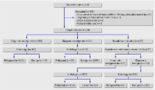
          Participants 1938 women with an adnexal mass examined with ultrasound by the principal investigator at each centre with a standardised research protocol.

          Reference standard Histological classification of the excised adnexal mass as benign or malignant.

          Main outcome measures Diagnostic sensitivity and specificity.

          Results Of the 1938 patients with an adnexal mass, 1396 (72%) had benign tumours, 373 (19.2%) had primary invasive tumours, 111 (5.7%) had borderline malignant tumours, and 58 (3%) had metastatic tumours in the ovary. The simple rules yielded a conclusive result in 1501 (77%) masses, for which they resulted in a sensitivity of 92% (95% confidence interval 89% to 94%) and a specificity of 96% (94% to 97%). The corresponding sensitivity and specificity of subjective assessment were 91% (88% to 94%) and 96% (94% to 97%). In the 357 masses for which the simple rules yielded an inconclusive result and with available results of CA-125 measurements, the sensitivities were 89% (83% to 93%) for subjective assessment, 50% (42% to 58%) for the risk of malignancy index, 89% (83% to 93%) for logistic regression model 1, and 82% (75% to 87%) for logistic regression model 2; the corresponding specificities were 78% (72% to 83%), 84% (78% to 88%), 44% (38% to 51%), and 48% (42% to 55%). Use of the simple rules as a triage test and subjective assessment for those masses for which the simple rules yielded an inconclusive result gave a sensitivity of 91% (88% to 93%) and a specificity of 93% (91% to 94%), compared with a sensitivity of 90% (88% to 93%) and a specificity of 93% (91% to 94%) when subjective assessment was used in all masses.

          Conclusions The use of the simple rules has the potential to improve the management of women with adnexal masses. In adnexal masses for which the rules yielded an inconclusive result, subjective assessment of ultrasonic findings by an experienced ultrasound examiner was the most accurate diagnostic test; the risk of malignancy index and the two regression models were not useful.

          Related collections

          Most cited references24

          • Record: found
          • Abstract: not found
          • Article: not found

          Carcinoma of the ovary. FIGO 26th Annual Report on the Results of Treatment in Gynecological Cancer.

            Bookmark
            • Record: found
            • Abstract: not found
            • Article: not found

            Prognosis and prognostic research: validating a prognostic model.

              Bookmark
              • Record: found
              • Abstract: not found
              • Article: not found

              Terms, definitions and measurements to describe the sonographic features of adnexal tumors: a consensus opinion from the International Ovarian Tumor Analysis (IOTA) Group.

                Bookmark

                Author and article information

                Contributors
                Role: professor in obstetrics and gynaecology
                Role: postdoctoral researcher in biostatistics
                Role: consultant gynaecologist
                Role: associate professor in obstetrics and gynaecology
                Role: professor in obstetrics and gynaecology
                Role: associate professor in obstetrics and gynaecology
                Role: consultant gynaecologist
                Role: consultant gynaecologist
                Role: consultant gynaecologist
                Role: consultant gynaecologist
                Role: assistant professor in gynaecology
                Role: research fellow in gynaecology
                Role: professor in obstetrics and gynaecology
                Role: professor in biomedical data processing
                Role: consultant gynaecologist, Role: visiting professor in obstetrics and gynaecology
                Role: professor in obstetrics and gynaecology
                Journal
                BMJ
                bmj
                BMJ : British Medical Journal
                BMJ Publishing Group Ltd.
                0959-8138
                1468-5833
                2010
                2010
                14 December 2010
                : 341
                : c6839
                Affiliations
                [1 ]Department of Obstetrics and Gynaecology, University Hospitals KU Leuven, 3000 Leuven, Belgium
                [2 ]Department of Electrical Engineering, ESAT-SCD, Katholieke Universiteit Leuven, 3001 Heverlee-Leuven, Belgium
                [3 ]Oncogynaecological Center, Department of Obstetrics and Gynecology, First Faculty of Medicine and General University Hospital of Charles University, 128 08 Prague 2, Czech Republic
                [4 ]Department of Obstetrics and Gynaecology, Lund University, 22185 Lund, Sweden
                [5 ]Department of Obstetrics and Gynaecology, University of Cagliari, Ospedale San Giovanni di Dio, 09124 Cagliari, Sardinia, Italy
                [6 ]Department of Obstetrics and Gynaecology, Ziekenhuis Oost-Limburg, Genk (ZOL) 3600 Genk, Belgium
                [7 ]Reproductive Medicine Unit, Department of Obstetrics and Gynaecology, University of Bologna, 40138 Bologna, Italy
                [8 ]Clinica Ostetrica e Ginecologica, Ospedale S Gerardo, Università di Milano Bicocca, Monza I-20052, Italy
                [9 ]Istituto di Clinica Ostetrica e Ginecologica, Università Cattolica di Sacro Cuore, 00168 Roma, Italy
                [10 ]Department of Obstetrics and Gynaecology, Imperial College London, Queen Charlotte’s and Chelsea Hospital, London W12 0HS, UK
                [11 ]Department of Obstetrics and Gynaecology, Malmö University Hospital, Lund University, SE20502 Malmö, Sweden
                Author notes
                Correspondence to: D Timmerman dirk.timmerman@ 123456uzleuven.be
                Article
                timd784744
                10.1136/bmj.c6839
                3001703
                21156740
                db459ba0-441e-47ef-84fb-2e3fdf7cdebd
                © Timmerman et al 2010

                This is an open-access article distributed under the terms of the Creative Commons Attribution Non-commercial License, which permits use, distribution, and reproduction in any medium, provided the original work is properly cited, the use is non commercial and is otherwise in compliance with the license. See: http://creativecommons.org/licenses/by-nc/2.0/ and http://creativecommons.org/licenses/by-nc/2.0/legalcode.

                History
                : 29 September 2010
                Categories
                Research
                Reproductive Medicine
                Screening (Oncology)
                Radiology
                Clinical Diagnostic Tests
                Surgical Oncology

                Medicine
                Medicine

                Comments

                Comment on this article