8
views
0
recommends
+1 Recommend
0 collections
    0
    shares
      • Record: found
      • Abstract: found
      • Article: found
      Is Open Access

      Algorithm for Correcting the Keratometric Error in the Estimation of the Corneal Power in Keratoconus Eyes after Accelerated Corneal Collagen Crosslinking

      research-article

      Read this article at

      Bookmark
          There is no author summary for this article yet. Authors can add summaries to their articles on ScienceOpen to make them more accessible to a non-specialist audience.

          Abstract

          Purpose

          To analyze the errors associated to corneal power calculation using the keratometric approach in keratoconus eyes after accelerated corneal collagen crosslinking (CXL) surgery and to obtain a model for the estimation of an adjusted corneal refractive index ( n k adj ) minimizing such errors.

          Methods

          Potential differences (Δ P c) among keratometric ( P k ) and Gaussian corneal power ( P c Gauss) were simulated. Three algorithms based on the use of n k adj for the estimation of an adjusted keratometric corneal power ( P k adj ) were developed. The agreement between P k(1.3375) (keratometric power using the keratometric index of 1.3375), P c Gauss, and P kadj was evaluated. The validity of the algorithm developed was investigated in 21 keratoconus eyes undergoing accelerated CXL.

          Results

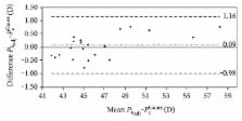
          P k(1.3375) overestimated corneal power between 0.3 and 3.2 D in theoretical simulations and between 0.8 and 2.9 D in the clinical study (Δ P c). Three linear equations were defined for n k adj to be used for different ranges of r 1c. In the clinical study, differences between P k adj and P c Gauss did not exceed ±0.8 D n k = 1.3375. No statistically significant differences were found between P k adj and P c Gauss ( p > 0.05) and P k(1.3375) and P k adj ( p < 0.001).

          Conclusions

          The use of the keratometric approach in keratoconus eyes after accelerated CXL can lead to significant clinical errors. These errors can be minimized with an adjusted keratometric approach.

          Related collections

          Most cited references17

          • Record: found
          • Abstract: found
          • Article: not found

          Corneal collagen crosslinking using riboflavin and ultraviolet-A light for keratoconus: one-year analysis using Scheimpflug imaging.

          To evaluate changes in corneal curvature, corneal elevation, corneal thickness, lens density, and foveal thickness after corneal collagen crosslinking with riboflavin and ultraviolet-A (UVA) light in eyes with progressive keratoconus. Grewal Eye Institute, Chandigarh, India. Subjective refraction, best corrected visual acuity (BCVA), Scheimpflug imaging, and optical coherence tomography were performed preoperatively and 1 week, 1, 3, and 6 months, and 1 year after crosslinking. There were no significant differences (P > 0.05) in mean values between preoperatively and 1 year postoperatively, respectively, in BCVA (0.22 +/- 0.10 and 0.20 +/- 0.10), spherical equivalent (-6.30 +/- 4.50 diopters (D) and -4.90 +/- 3.50 D), or cylinder vector (1.58 x 7( degrees ) +/- 3.8 D and 1.41 x 24( degrees ) +/- 3.5 D). There was no significant difference in mean measurements between preoperatively and 1 year postoperatively, respectively, for central corneal thickness (458.9 +/- 40 microm and 455.2 +/- 48.6 microm), anterior corneal curvature (50.6 +/- 7.4 D and 51.5 +/- 3.6 D), posterior corneal curvature (-7.7 +/- 1.2 D and -7.4 +/- 1.1 D), apex anterior (P = .9), posterior corneal elevation (P = .7), lens density (P = .33), or foveal thickness (175.7 +/- 35.6 microm and 146.4 +/- 8.5 microm; P = .1). Stable BCVA, spherical equivalent, anterior and posterior corneal curvatures, and corneal elevation 1 year after crosslinking indicate that keratoconus did not progress. Unchanged lens density and foveal thickness suggest that the lens and macula were not affected after UVA exposure during crosslinking.
            Bookmark
            • Record: found
            • Abstract: not found
            • Article: not found

            Statistics Notes: Measurement error and correlation coefficients

              Bookmark
              • Record: found
              • Abstract: found
              • Article: not found

              Longitudinal study of keratoconus progression.

              To determine if differences in topographic progression between unaffected keratoconus relatives and normal controls can predict factors associated with the development of keratoconus in a longitudinal study. We recruited 369 unaffected keratoconus relatives and 119 normal controls in Los Angeles. Both eyes of subjects were examined at baseline clinically and by quantitative videokeratography and at a period ranging from 1 year to 8 years. Progression to keratoconus was evaluated by quantitative videokeratography variables. Unaffected relatives had higher Central K (CK), I-S and KISA values and were younger than normal controls (CK: 44.70 vs 44.01, P or = 47.2 and I-S> or =1.2 or KISA> or = 60) and low risk (age>30 and Central K<47.2 and I-S<1.2 and KISA< 60), relatives in the high risk group had a greater increase in CK and I-S values than those in the low risk group (CK: P=0.009; I-S: P<0.001), which indicated that there were significantly different rates of progression between two groups. Unaffected relatives had higher videokeratography indices than normal controls, but overall they did not progress to keratoconus quicker than normal controls. However, relatives in the high risk group may have a greater risk of progression to keratoconus.
                Bookmark

                Author and article information

                Contributors
                Journal
                J Ophthalmol
                J Ophthalmol
                JOPH
                Journal of Ophthalmology
                Hindawi
                2090-004X
                2090-0058
                2017
                22 October 2017
                : 2017
                : 8529489
                Affiliations
                1Grupo de Óptica y Percepción Visual (GOPV), Department of Optics, Pharmacology and Anatomy, University of Alicante, Alicante, Spain
                2Department of Ophthalmology (OFTALMAR), Medimar International Hospital, Alicante, Spain
                3Fundación para la Calidad Visual (FUNCAVIS), Alicante, Spain
                Author notes

                Academic Editor: Edward Manche

                Author information
                http://orcid.org/0000-0002-1546-4807
                http://orcid.org/0000-0003-2150-3830
                Article
                10.1155/2017/8529489
                5672131
                db2903ec-d3d7-4814-a113-a3fbffbe5297
                Copyright © 2017 David P. Piñero et al.

                This is an open access article distributed under the Creative Commons Attribution License, which permits unrestricted use, distribution, and reproduction in any medium, provided the original work is properly cited.

                History
                : 18 June 2017
                : 19 September 2017
                Categories
                Research Article

                Ophthalmology & Optometry
                Ophthalmology & Optometry

                Comments

                Comment on this article