28
views
0
recommends
+1 Recommend
1 collections
    0
    shares
      • Record: found
      • Abstract: found
      • Article: not found

      Exploring the Effect of Image Enhancement Techniques on COVID-19 Detection using Chest X-rays Images

      research-article

      Read this article at

      ScienceOpenPublisherPMC
      Bookmark
          There is no author summary for this article yet. Authors can add summaries to their articles on ScienceOpen to make them more accessible to a non-specialist audience.

          Abstract

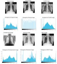
          Computer-aided diagnosis for the reliable and fast detection of coronavirus disease (COVID-19) has become a necessity to prevent the spread of the virus during the pandemic to ease the burden on the healthcare system. Chest X-ray (CXR) imaging has several advantages over other imaging and detection techniques. Numerous works have been reported on COVID-19 detection from a smaller set of original X-ray images. However, the effect of image enhancement and lung segmentation of a large dataset in COVID-19 detection was not reported in the literature. We have compiled a large X-ray dataset (COVQU) consisting of 18,479 CXR images with 8851 normal, 6012 non-COVID lung infections, and 3616 COVID-19 CXR images and their corresponding ground truth lung masks. To the best of our knowledge, this is the largest public COVID positive database and the lung masks. Five different image enhancement techniques: histogram equalization (HE), contrast limited adaptive histogram equalization (CLAHE), image complement, gamma correction, and balance contrast enhancement technique (BCET) were used to investigate the effect of image enhancement techniques on COVID-19 detection. A novel U-Net model was proposed and compared with the standard U-Net model for lung segmentation. Six different pre-trained Convolutional Neural Networks (CNNs) (ResNet18, ResNet50, ResNet101, InceptionV3, DenseNet201, and ChexNet) and a shallow CNN model were investigated on the plain and segmented lung CXR images. The novel U-Net model showed an accuracy, Intersection over Union (IoU), and Dice coefficient of 98.63%, 94.3%, and 96.94%, respectively for lung segmentation. The gamma correction-based enhancement technique outperforms other techniques in detecting COVID-19 from the plain and the segmented lung CXR images. Classification performance from plain CXR images is slightly better than the segmented lung CXR images; however, the reliability of network performance is significantly improved for the segmented lung images, which was observed using the visualization technique. The accuracy, precision, sensitivity, F1-score, and specificity were 95.11 %, 94.55 %, 94.56 %, 94.53 %, and 95.59 % respectively for the segmented lung images. The proposed approach with very reliable and comparable performance will boost the fast and robust COVID-19 detection using chest X-ray images.

          Related collections

          Most cited references19

          • Record: found
          • Abstract: found
          • Article: not found

          Covid-19: automatic detection from X-ray images utilizing transfer learning with convolutional neural networks

          In this study, a dataset of X-ray images from patients with common bacterial pneumonia, confirmed Covid-19 disease, and normal incidents, was utilized for the automatic detection of the Coronavirus disease. The aim of the study is to evaluate the performance of state-of-the-art convolutional neural network architectures proposed over the recent years for medical image classification. Specifically, the procedure called Transfer Learning was adopted. With transfer learning, the detection of various abnormalities in small medical image datasets is an achievable target, often yielding remarkable results. The datasets utilized in this experiment are two. Firstly, a collection of 1427 X-ray images including 224 images with confirmed Covid-19 disease, 700 images with confirmed common bacterial pneumonia, and 504 images of normal conditions. Secondly, a dataset including 224 images with confirmed Covid-19 disease, 714 images with confirmed bacterial and viral pneumonia, and 504 images of normal conditions. The data was collected from the available X-ray images on public medical repositories. The results suggest that Deep Learning with X-ray imaging may extract significant biomarkers related to the Covid-19 disease, while the best accuracy, sensitivity, and specificity obtained is 96.78%, 98.66%, and 96.46% respectively. Since by now, all diagnostic tests show failure rates such as to raise concerns, the probability of incorporating X-rays into the diagnosis of the disease could be assessed by the medical community, based on the findings, while more research to evaluate the X-ray approach from different aspects may be conducted.
            Bookmark
            • Record: found
            • Abstract: found
            • Article: not found

            Convolutional Neural Networks for Medical Image Analysis: Full Training or Fine Tuning?

            Training a deep convolutional neural network (CNN) from scratch is difficult because it requires a large amount of labeled training data and a great deal of expertise to ensure proper convergence. A promising alternative is to fine-tune a CNN that has been pre-trained using, for instance, a large set of labeled natural images. However, the substantial differences between natural and medical images may advise against such knowledge transfer. In this paper, we seek to answer the following central question in the context of medical image analysis: Can the use of pre-trained deep CNNs with sufficient fine-tuning eliminate the need for training a deep CNN from scratch? To address this question, we considered four distinct medical imaging applications in three specialties (radiology, cardiology, and gastroenterology) involving classification, detection, and segmentation from three different imaging modalities, and investigated how the performance of deep CNNs trained from scratch compared with the pre-trained CNNs fine-tuned in a layer-wise manner. Our experiments consistently demonstrated that 1) the use of a pre-trained CNN with adequate fine-tuning outperformed or, in the worst case, performed as well as a CNN trained from scratch; 2) fine-tuned CNNs were more robust to the size of training sets than CNNs trained from scratch; 3) neither shallow tuning nor deep tuning was the optimal choice for a particular application; and 4) our layer-wise fine-tuning scheme could offer a practical way to reach the best performance for the application at hand based on the amount of available data.
              Bookmark
              • Record: found
              • Abstract: found
              • Article: found
              Is Open Access

              COVID-Net: a tailored deep convolutional neural network design for detection of COVID-19 cases from chest X-ray images

              The Coronavirus Disease 2019 (COVID-19) pandemic continues to have a devastating effect on the health and well-being of the global population. A critical step in the fight against COVID-19 is effective screening of infected patients, with one of the key screening approaches being radiology examination using chest radiography. It was found in early studies that patients present abnormalities in chest radiography images that are characteristic of those infected with COVID-19. Motivated by this and inspired by the open source efforts of the research community, in this study we introduce COVID-Net, a deep convolutional neural network design tailored for the detection of COVID-19 cases from chest X-ray (CXR) images that is open source and available to the general public. To the best of the authors’ knowledge, COVID-Net is one of the first open source network designs for COVID-19 detection from CXR images at the time of initial release. We also introduce COVIDx, an open access benchmark dataset that we generated comprising of 13,975 CXR images across 13,870 patient patient cases, with the largest number of publicly available COVID-19 positive cases to the best of the authors’ knowledge. Furthermore, we investigate how COVID-Net makes predictions using an explainability method in an attempt to not only gain deeper insights into critical factors associated with COVID cases, which can aid clinicians in improved screening, but also audit COVID-Net in a responsible and transparent manner to validate that it is making decisions based on relevant information from the CXR images. By no means a production-ready solution, the hope is that the open access COVID-Net, along with the description on constructing the open source COVIDx dataset, will be leveraged and build upon by both researchers and citizen data scientists alike to accelerate the development of highly accurate yet practical deep learning solutions for detecting COVID-19 cases and accelerate treatment of those who need it the most.
                Bookmark

                Author and article information

                Journal
                Comput Biol Med
                Comput Biol Med
                Computers in Biology and Medicine
                Elsevier Ltd.
                0010-4825
                1879-0534
                11 March 2021
                11 March 2021
                : 104319
                Affiliations
                [a ]Department of Biomedical Physics & Technology, University of Dhaka Dhaka, 1000, Bangladesh
                [b ]Department of Electrical Engineering, Qatar University, Doha, 2713, Qatar
                [c ]Faculty of Robotics and Advanced Computing, Qatar Armed Forces Academic Bridge Program, Qatar Foundation, Doha, 24404, Qatar
                [d ]Dept. of Electrical, Electronics and Systems Engineering, Universiti Kebangsaan Malaysia, Bangi, Selangor, 43600, Malaysia
                [e ]Department of Computer Science and Engineering, Qatar University, Doha, 2713, Qatar
                [f ]Department of Basic Medical Sciences, College of Medicine, Biomedical and Pharmaceutical Research Unit, QU Health, Qatar University, Doha, 2713, Qatar
                [g ]Department of Electrical Engineering (JC), University of Engineering and Technology, Peshawar, Pakistan
                Author notes
                []Corresponding author.
                Article
                S0010-4825(21)00113-X 104319
                10.1016/j.compbiomed.2021.104319
                7946571
                33799220
                db02f43d-e685-485c-a461-35baa1b11579
                © 2021 Elsevier Ltd. All rights reserved.

                Since January 2020 Elsevier has created a COVID-19 resource centre with free information in English and Mandarin on the novel coronavirus COVID-19. The COVID-19 resource centre is hosted on Elsevier Connect, the company's public news and information website. Elsevier hereby grants permission to make all its COVID-19-related research that is available on the COVID-19 resource centre - including this research content - immediately available in PubMed Central and other publicly funded repositories, such as the WHO COVID database with rights for unrestricted research re-use and analyses in any form or by any means with acknowledgement of the original source. These permissions are granted for free by Elsevier for as long as the COVID-19 resource centre remains active.

                History
                : 24 November 2020
                : 3 March 2021
                : 4 March 2021
                Categories
                Article

                covid-19,image enhancement,chest x-ray images,convolutional neural networks,lung segmentation

                Comments

                Comment on this article