0
views
0
recommends
+1 Recommend
0 collections
    0
    shares
      • Record: found
      • Abstract: found
      • Article: found
      Is Open Access

      Inflammatory Myofibroblastic Tumor of the Sciatic Nerve Mimicking Lumbar Disc Herniation: A Diagnostic Challenge

      case-report

      Read this article at

      Bookmark
          There is no author summary for this article yet. Authors can add summaries to their articles on ScienceOpen to make them more accessible to a non-specialist audience.

          Abstract

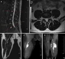
          Inflammatory myofibroblastic tumors (IMTs), which involve the proliferation of fibroblastic-myofibroblastic cells mixed with inflammatory infiltrates, are exceedingly rare in the extremities. There are no reported IMTs involving the sciatic nerve. This type of involvement may cause entrapment of the sciatic nerve, whose symptoms may mimic lumbar disc herniation (LDH), especially when it occurs in patients with lumbar degenerative disc disease. We describe the case of a 40-year-old male with lumbar degenerative disc disease accompanied by IMT involving the sciatic nerve whose symptoms mimicked LDH and posed a diagnostic challenge. We showed the course of the disease as well as the systematic imaging manifestations of IMTs involving the sciatic nerve and discussed their therapeutic management.

          Related collections

          Most cited references21

          • Record: found
          • Abstract: found
          • Article: not found

          Inflammatory myofibroblastic tumor: comparison of clinicopathologic, histologic, and immunohistochemical features including ALK expression in atypical and aggressive cases.

          Inflammatory myofibroblastic tumor (IMT) is a neoplasm of intermediate biologic potential. In this study, we report a subset of IMTs with histologic atypia and/or clinical aggressiveness that were analyzed for clinicopathologic features, outcome, and immunohistochemical expression of anaplastic lymphoma kinase (ALK) and other markers to identify potential pathologic prognostic features. Fifty-nine IMTs with classic morphology (5 cases), atypical histologic features (21 cases), local recurrence (27 cases), and/or metastasis (6 cases) were studied. Immunohistochemistry was performed for ALK1 and other markers (Mib-1, c-Myc, cyclin D1, caspase 3, Bcl-2, Mcl-1, survivin, p27, CD56, p53, MDM-2) using standard techniques. The 59 IMTs had an age at diagnosis ranging from 3 weeks to 74 years (mean 13.2 y, median 11 y, 44% in the first decade). The mean tumor size was 7.8 cm. Sites included the abdomen or pelvis in 64%, lung in 22%, head and neck in 8%, and extremities in 5%. The follow-up ranged from 3 months to 11 years, with a mean of 3.6 years and a median of 3 years. Thirty-three patients had local recurrences, including 13 with multiple local recurrences and 6 patients with both local recurrences and distant metastases. Six patients died of disease, 5 with local recurrences, and 1 with distant metastases. Histologic evolution to a more pleomorphic cellular, spindled, polygonal, or round cell morphologic pattern was observed in 7 cases. Abdominal and pelvic IMTs had a recurrence rate of 85%. Recurrent and metastatic IMTs were larger, with mean diameters of 8.7 and 11 cm, respectively. Cytoplasmic ALK reactivity was seen in 56%. ALK-negative IMTs occurred in older patients (mean age 20.1) years and had greater nuclear pleomorphism, atypia, and atypical mitoses. All 6 metastatic IMTs were ALK-negative. Nuclear expression of p53 was detected in 80% of IMTs overall, but in only 25% of the metastatic subset. There were no significant differences among the subgroups for c-Myc, cyclin D1, MDM-2, Mcl-1, Bcl-2, CD56, p27, caspase 3, or survivin expression. In conclusion, among these 59 IMTs, ALK reactivity was associated with local recurrence, but not distant metastasis, which was confined to ALK-negative lesions. Absent ALK expression was associated with a higher age overall, subtle histologic differences, and death from disease or distant metastases (in a younger subset). Other proliferative, apoptotic, and prognostic markers did not correlate well with morphology or outcome. Thus, ALK reactivity may be a favorable prognostic indicator in IMT and abdominopelvic IMTs recur more frequently.
            Bookmark
            • Record: found
            • Abstract: found
            • Article: not found

            Targeting ALK With Crizotinib in Pediatric Anaplastic Large Cell Lymphoma and Inflammatory Myofibroblastic Tumor: A Children's Oncology Group Study.

            Purpose Fusions involving the ALK gene are the predominant genetic lesion underlying pediatric anaplastic large cell lymphomas (ALCL) and inflammatory myofibroblastic tumors (IMTs). We assessed the activity of the ALK inhibitor crizotinib in patients who had no known curative treatment options at diagnosis or with relapsed/recurrent disease. Methods In this study, 26 patients with relapsed/refractory ALK-positive ALCL and 14 patients with metastatic or inoperable ALK-positive IMT received crizotinib orally twice daily. Study objectives were measurement of efficacy and safety. Correlative studies evaluated the serial detection of NPM-ALK fusion transcripts in patients with ALCL. Results The overall response rates for patients with ALCL treated at doses of 165 (ALCL165) and 280 (ALCL280) mg/m(2) were 83% and 90%, respectively. The overall response rate for patients with IMT (treated at 100, 165, and 280 mg/m(2)/dose) was 86%. A complete response was observed in 83% (five of six) of ALCL165, 80% (16 of 20) of ALCL280, and 36% (five of 14) of patients with IMT. Partial response rates were 0% (none of six), 10% (two of 20), and 50% (seven of 14), respectively. The median duration of therapy was 2.79, 0.4, and 1.63 years, respectively, with 12 patients ceasing protocol therapy to proceed to transplantation. The most common drug-related adverse event was decrease in neutrophil count in 33% and 70% of the ALCL165 and ALCL280 groups, respectively, and in 43% of patients with IMT. Levels of NPM-ALK decreased during therapy in most patients with ALCL. Conclusion The robust and sustained clinical responses to crizotinib therapy in patients with relapsed ALCL and metastatic or unresectable IMT highlight the importance of the ALK pathway in these diseases.
              Bookmark
              • Record: found
              • Abstract: found
              • Article: not found

              Inflammatory myofibroblastic tumors (inflammatory pseudotumors) of the gastrointestinal tract: how closely are they related to inflammatory fibroid polyps?

              Inflammatory myofibroblastic tumors (inflammatory pseudotumors) and inflammatory fibroid polyps of the gastrointestinal tract both feature prominent inflammatory infiltrates admixed with spindle-shaped fibroblasts/myofibroblasts set in a collagenous, fibrovascular, or myxoid stroma. Erroneously, some have considered inflammatory fibroid polyps to be intraluminal manifestations of inflammatory myofibroblastic tumors. In this study, we have characterized the histopathology of inflammatory myofibroblastic tumors, tumors that have only rarely been reported in the gastrointestinal tract, and have focused on whether inflammatory myofibroblastic tumors and inflammatory fibroid polyps in the gastrointestinal tract are distinct or similar. Clinical, histopathologic, and immunohistochemical features of 38 inflammatory myofibroblastic tumors limited to the wall of the gastrointestinal tract were compared with those of 45 inflammatory fibroid polyps. Compared to patients with inflammatory fibroid polyps, those with inflammatory myofibroblastic tumors were younger (mean age 41 years vs. 53 years); had larger tumors (mean 8 +/- 5.2 cm vs. 3.6 +/- 4.6 cm); presented with abdominal pain, fever, and weight loss more frequently and less frequently had bowel obstruction. Inflammatory fibroid polyps had more eosinophils and fibrosis and fewer lymphoid cell infiltrates than inflammatory myofibroblastic tumors. A regular vascular pattern was a feature of inflammatory fibroid polyps but not of inflammatory myofibroblastic tumors. Most (82%) inflammatory fibroid polyps were positive for CD34 versus none of the inflammatory myofibroblastic tumors. Smooth muscle actin was more frequently positive in inflammatory myofibroblastic tumors than in inflammatory fibroid polyps (86% versus 13%). Inflammatory myofibroblastic tumors were much less frequent and were more evenly distributed in the gastrointestinal tract than inflammatory fibroid polyps. Both appear to be benign processes. Inflammatory myofibroblastic tumors, but not inflammatory fibroid polyps, had a tendency to recur. In conclusion, inflammatory myofibroblastic tumors of the gastrointestinal tract are extremely rare and differ clinically, histologically, and immunohistochemically from inflammatory fibroid polyps. Copyright 2002, Elsevier Science (USA). All rights reserved.
                Bookmark

                Author and article information

                Journal
                World J Oncol
                World J Oncol
                Elmer Press
                World Journal of Oncology
                Elmer Press
                1920-4531
                1920-454X
                June 2024
                15 April 2024
                : 15
                : 3
                : 521-525
                Affiliations
                [a ]Department of Orthopedic Surgery, The First Affiliated Hospital of Shandong First Medical University & Shandong Provincial Qianfoshan Hospital, Shandong Key Laboratory of Rheumatic Disease and Translational Medicine, Jinan, China
                [b ]International Medical Center, Affiliated Hospital of Qingdao University Medical College, Qingdao, China
                [c ]Department of Orthopedic Surgery, Shandong Provincial Hospital Affiliated to Shandong First Medical University, Jinan, China
                [d ]Chang Jun Chen and Jun Feng Yin contributed equally to this work.
                Author notes
                [e ]Corresponding Author: Chang Jun Chen and Da Yong Peng, Department of Orthopedic Surgery, The First Affiliated Hospital of Shandong First Medical University & Shandong Provincial Qianfoshan Hospital, Shandong Key Laboratory of Rheumatic Disease and Translational Medicine, Jinan, China. Email: Chenchangjun888@ 123456qq.com and 979837284@ 123456qq.com
                Author information
                https://orcid.org/0000-0002-4198-7177
                Article
                10.14740/wjon1777
                11092406
                38751705
                daf0b66f-6c39-4762-89be-8b0d8c4e13d1
                Copyright 2024, Chen et al.

                This article is distributed under the terms of the Creative Commons Attribution Non-Commercial 4.0 International License, which permits unrestricted non-commercial use, distribution, and reproduction in any medium, provided the original work is properly cited.

                History
                : 28 November 2023
                : 16 March 2024
                Funding
                This work was supported by the Natural Science Foundation of Shandong Province (grant no. ZR2023QH517; ZR2022QH252); Clinical Medical Science and Technology Innovation Program of Jinan Science and Technology Bureau (grant no. 202328059); Foundation of Research Hospital Association of Shandong Province (grant no. 2022016); Foundation of Scientific Research Incubation of Shandong First Medical University and Shandong Academy of Medical Sciences (grant no. 202201-086); Taishan Scholar Program of Shandong Province (No.tsqn202312357); National Science Foundation of China (No.81902188).
                Categories
                Case Report

                inflammatory myofibroblastic tumor,sciatic nerve,lumbar disc herniation

                Comments

                Comment on this article