19
views
0
recommends
+1 Recommend
0 collections
    0
    shares
      • Record: found
      • Abstract: found
      • Article: found
      Is Open Access

      Extracellular sphingomyelinase activity impairs TNF-α-induced endothelial cell death via ADAM17 activation and TNF receptor 1 shedding

      research-article

      Read this article at

      Bookmark
          There is no author summary for this article yet. Authors can add summaries to their articles on ScienceOpen to make them more accessible to a non-specialist audience.

          Abstract

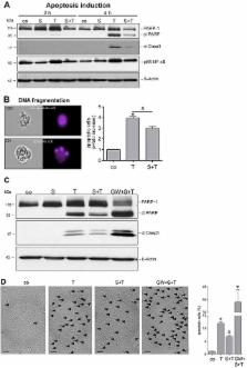
          ADAM17, a prominent member of the “Disintegrin and Metalloproteinase” (ADAM) family, is an important regulator of endothelial cell proliferation and cell survival. The protease controls vital cellular functions through cleavage of growth factors, cytokines and their receptors including transforming growth factor-alpha (TGF-α), tumor necrosis factor-alpha (TNF-α) and TNF-α receptor 1 (TNFR1). TNF-α is the major inducer of endothelial cell death in cardiovascular diseases. The latter are also characterized by elevated plasma and tissue levels of extracellular sphingomyelinase (SMase). Whether the SMase affects ADAM activity and thus endothelial cell function has not been addressed to date. Here, we analyzed the effect of SMase on ADAM17-mediated shedding in COS7 cells and in human umbilical vein endothelial cells (HUVECs). Exposure to SMase significantly increased ADAM17-mediated release of alkaline-phosphatase (AP)-tagged TGF-α in COS7 cells and shedding of endogenously expressed TNFR1 in HUVECs. We previously presented evidence that surface exposure of phosphatidylserine (PS) is pivotal for ADAM17 to exert sheddase function. We found that SMase treatment led to PS externalization in both cell types. Transient non-apoptotic PS exposure is often mediated by Ca 2+-dependent phospholipid scramblases. Accordingly, the Ca 2+-chelator EGTA markedly reduced the breakdown of phospholipid asymmetry and shedding of TGF-α and TNFR1. Moreover, sheddase activity was significantly diminished in the presence of the competing PS-headgroup OPLS. SMase-stimulated TNFR1 shedding strikingly diminished TNF-α-induced signalling cascades and endothelial cell death. Taken together, our data suggest that SMase activity might act as protective factor for endothelial cells in cardiovascular diseases.

          Related collections

          Most cited references40

          • Record: found
          • Abstract: found
          • Article: not found

          Calcium-dependent phospholipid scrambling by TMEM16F.

          In all animal cells, phospholipids are asymmetrically distributed between the outer and inner leaflets of the plasma membrane. This asymmetrical phospholipid distribution is disrupted in various biological systems. For example, when blood platelets are activated, they expose phosphatidylserine (PtdSer) to trigger the clotting system. The PtdSer exposure is believed to be mediated by Ca(2+)-dependent phospholipid scramblases that transport phospholipids bidirectionally, but its molecular mechanism is still unknown. Here we show that TMEM16F (transmembrane protein 16F) is an essential component for the Ca(2+)-dependent exposure of PtdSer on the cell surface. When a mouse B-cell line, Ba/F3, was treated with a Ca(2+) ionophore under low-Ca(2+) conditions, it reversibly exposed PtdSer. Using this property, we established a Ba/F3 subline that strongly exposed PtdSer by repetitive fluorescence-activated cell sorting. A complementary DNA library was constructed from the subline, and a cDNA that caused Ba/F3 to expose PtdSer spontaneously was identified by expression cloning. The cDNA encoded a constitutively active mutant of TMEM16F, a protein with eight transmembrane segments. Wild-type TMEM16F was localized on the plasma membrane and conferred Ca(2+)-dependent scrambling of phospholipids. A patient with Scott syndrome, which results from a defect in phospholipid scrambling activity, was found to carry a mutation at a splice-acceptor site of the gene encoding TMEM16F, causing the premature termination of the protein.
            Bookmark
            • Record: found
            • Abstract: found
            • Article: not found

            Endothelial apoptosis as the primary lesion initiating intestinal radiation damage in mice.

            Gastrointestinal (GI) tract damage by chemotherapy or radiation limits their efficacy in cancer treatment. Radiation has been postulated to target epithelial stem cells within the crypts of Lieberkühn to initiate the lethal GI syndrome. Here, we show in mouse models that microvascular endothelial apoptosis is the primary lesion leading to stem cell dysfunction. Radiation-induced crypt damage, organ failure, and death from the GI syndrome were prevented when endothelial apoptosis was inhibited pharmacologically by intravenous basic fibroblast growth factor (bFGF) or genetically by deletion of the acid sphingomyelinase gene. Endothelial, but not crypt, cells express FGF receptor transcripts, suggesting that the endothelial lesion occurs before crypt stem cell damage in the evolution of the GI syndrome. This study provides a basis for new approaches to prevent radiation damage to the bowel.
              Bookmark
              • Record: found
              • Abstract: found
              • Article: not found

              Functions of ceramide in coordinating cellular responses to stress.

              Y Hannun (1996)
              Sphingolipid metabolites participate in key events of signal transduction and cell regulation. In the sphingomyelin cycle, a number of extracellular agents and insults (such as tumor necrosis factor, Fas ligands, and chemotherapeutic agents) cause the activation of sphingomyelinases, which act on membrane sphingomyelin and release ceramide. Multiple experimental approaches suggest an important role for ceramide in regulating such diverse responses as cell cycle arrest, apoptosis, and cell senescence. In vitro, ceramide activates a serine-threonine protein phosphatase, and in cells it regulates protein phosphorylation as well as multiple downstream targets [such as interleukin converting enzyme (ICE)-like proteases, stress-activated protein kinases, and the retinoblastoma gene product] that mediate its distinct cellular effects. This spectrum of inducers of ceramide accumulation and the nature of ceramide-mediated responses suggest that ceramide is a key component of intracellular stress response pathways.
                Bookmark

                Author and article information

                Journal
                Oncotarget
                Oncotarget
                Oncotarget
                ImpactJ
                Oncotarget
                Impact Journals LLC
                1949-2553
                22 September 2017
                7 August 2017
                : 8
                : 42
                : 72584-72596
                Affiliations
                1 Department of Dermatology, University of Kiel, 24105 Kiel, Germany
                2 Institute of Immunology, University of Kiel, 24105 Kiel, Germany
                Author notes
                Correspondence to: Karina Reiss, kreiss@ 123456dermatology.uni-kiel.de
                Article
                19983
                10.18632/oncotarget.19983
                5641154
                dae09a32-400c-4787-8541-4b8d6ba89785
                Copyright: © 2017 Sommer et al.

                This article is distributed under the terms of the Creative Commons Attribution License (CC-BY), which permits unrestricted use and redistribution provided that the original author and source are credited.

                History
                : 28 February 2017
                : 11 July 2017
                Categories
                Research Paper

                Oncology & Radiotherapy
                sphingomyelinase,adam17,apoptosis,tnf-α,tnfr1
                Oncology & Radiotherapy
                sphingomyelinase, adam17, apoptosis, tnf-α, tnfr1

                Comments

                Comment on this article