4
views
0
recommends
+1 Recommend
0 collections
    0
    shares
      • Record: found
      • Abstract: found
      • Article: found
      Is Open Access

      Determination and classification of intraoral phosphor storage plate artifacts and errors

      research-article

      Read this article at

      Bookmark
          There is no author summary for this article yet. Authors can add summaries to their articles on ScienceOpen to make them more accessible to a non-specialist audience.

          Abstract

          Purpose

          The aim of this study was to determine the reasons and solutions for intraoral phosphor storage plate (PSP) image artifacts and errors, and to develop an appropriate classification of the artifacts.

          Materials and Methods

          This study involved the retrospective examination of 5,000 intraoral images that had been obtained using a phosphor plate system. Image artifacts were examined on the radiographs and classified according to possible causative factors.

          Results

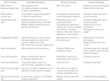
          Artifacts were observed in 1,822 of the 5,000 images. After examination of the images, the errors were divided into 6 groups based on their causes, as follows: images with operator errors, superposition of undesirable structures, ambient light errors, plate artifacts (physical deformations and contamination), scanner artifacts, and software artifacts. The groups were then re-examined and divided into 45 subheadings.

          Conclusion

          Identification of image artifacts can help to improve the quality of the radiographic image and control the radiation dose. Knowledge of the basic physics and technology of PSP systems could aid to reduce the need for repeated radiography.

          Related collections

          Most cited references12

          • Record: found
          • Abstract: found
          • Article: not found

          A method for the geometric and densitometric standardization of intraoral radiographs.

          The interpretation of dental radiographs for the diagnosis of periodontal disease conditions poses several difficulties. These include the inability to adequately reproduce the projection geometry and optical density of the exposures. In order to improve the ability to extract accurate quantitative information from a radiographic survey of periodontal status, a method was developed which provided for consistent reproduction of both geometric and densitometric exposure parameters. This technique employed vertical bitewing projections in holders customized to individual segments of the dentition. A copper stepwedge was designed to provide densitometric standardization, and wire markers were included to permit measurement of angular variation. In a series of 53 paired radiographs, measurement of alveolar crest heights was found to be reproducible within approximately 0.1 mm. This method provided a full mouth radiographic survey using seven films, each complete with internal standards suitable for computer-based image processing.
            Bookmark
            • Record: found
            • Abstract: found
            • Article: not found

            Effect of delayed scanning of storage phosphor plates.

            To test longevity of image quality in storage phosphor plates (SPPs) at various exposure settings, storage conditions, and delays in scanning.
              Bookmark
              • Record: found
              • Abstract: found
              • Article: not found

              Computed radiography image artifacts revisited.

              Computed radiography (CR) has provided a ready cost-effective transition from screen film to digital radiography and a convenient entrance to PACS. This article revisits artifacts encountered in CR systems. These artifacts may obscure abnormalities, mimic a clinical entity, or hamper image quality.
                Bookmark

                Author and article information

                Journal
                Imaging Sci Dent
                Imaging Sci Dent
                ISD
                Imaging Science in Dentistry
                Korean Academy of Oral and Maxillofacial Radiology
                2233-7822
                2233-7830
                September 2019
                24 September 2019
                : 49
                : 3
                : 219-228
                Affiliations
                [1 ]Department of Dental and Maxillofacial Radiology, Faculty of Dentistry, Çanakkale Onsekiz Mart University, Çanakkale, Turkey.
                [2 ]Department of Restorative Dentistry, Faculty of Dentistry, Alanya Alaaddin Keykubat University, Antalya, Turkey.
                Author notes
                Correspondence to: Prof. Yeşim Deniz. Department of Dental and Maxillofacial Radiology, Faculty of Dentistry, Çanakkale Onsekiz Mart University, 17110 Kepez, Çanakkale, Turkey. Tel) 90-507-3578000, yesimdeniz@ 123456comu.edu.tr
                Author information
                https://orcid.org/0000-0002-6967-5378
                https://orcid.org/0000-0003-2601-3064
                Article
                10.5624/isd.2019.49.3.219
                6761057
                31583205
                dab339bf-4c3d-4c20-a977-22e58b0038d2
                Copyright © 2019 by Korean Academy of Oral and Maxillofacial Radiology

                This is an Open Access article distributed under the terms of the Creative Commons Attribution Non-Commercial License ( http://creativecommons.org/licenses/by-nc/3.0/) which permits unrestricted non-commercial use, distribution, and reproduction in any medium, provided the original work is properly cited.

                History
                : 19 March 2019
                : 06 May 2019
                : 22 May 2019
                Categories
                Original Article

                Dentistry
                radiography, dental,diagnostic imaging,artifacts
                Dentistry
                radiography, dental, diagnostic imaging, artifacts

                Comments

                Comment on this article