14
views
0
recommends
+1 Recommend
1 collections
    0
    shares
      • Record: found
      • Abstract: found
      • Article: not found

      A SARS-CoV-2 Cluster in an Acute Care Hospital

      other

      Read this article at

      ScienceOpenPublisherPMC
      Bookmark
          There is no author summary for this article yet. Authors can add summaries to their articles on ScienceOpen to make them more accessible to a non-specialist audience.

          Abstract

          This study describes the detection, mitigation, and analysis of a large cluster of SARS-CoV-2 infections in an acute care hospital with mature infection control policies and discusses insights that may inform additional measures to protect patients and staff.

          Abstract

          Visual Abstract. SARS-CoV-2 Cluster in an Acute Care Hospital This study describes the detection, mitigation, and analysis of a large cluster of SARS-CoV-2 infections in an acute care hospital with mature infection control policies and discusses insights that may inform additional measures to protect patients and staff.
          Visual Abstract.
          SARS-CoV-2 Cluster in an Acute Care Hospital

          This study describes the detection, mitigation, and analysis of a large cluster of SARS-CoV-2 infections in an acute care hospital with mature infection control policies and discusses insights that may inform additional measures to protect patients and staff.

          Abstract

          Background:

          Little is known about clusters of severe acute respiratory syndrome coronavirus 2 (SARS-CoV-2) infection in acute care hospitals.

          Objective:

          To describe the detection, mitigation, and analysis of a large cluster of SARS-CoV-2 infections in an acute care hospital with mature infection control policies.

          Design:

          Descriptive study.

          Setting:

          Brigham and Women's Hospital, Boston, Massachusetts.

          Participants:

          Patients and staff with cluster-related SARS-CoV-2 infections.

          Intervention:

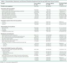
          Close contacts of infected patients and staff were identified and tested every 3 days, patients on affected units were preemptively isolated and repeatedly tested, affected units were cleaned, room ventilation was measured, and specimens were sent for whole-genome sequencing. A case–control study was done to compare clinical interactions, personal protective equipment use, and breakroom and workroom practices in SARS-CoV-2–positive versus negative staff.
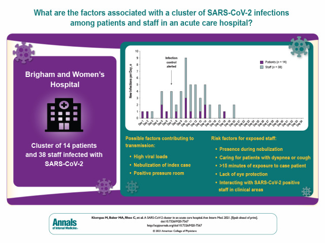
          Measurements:

          Description of the cluster, mitigation activities, and risk factor analysis.

          Results:

          Fourteen patients and 38 staff members were included in the cluster per whole-genome sequencing and epidemiologic associations. The index case was a symptomatic patient in whom isolation was discontinued after 2 negative results on nasopharyngeal polymerase chain reaction testing. The patient subsequently infected multiple roommates and staff, who then infected others. Seven of 52 (13%) secondary infections were detected only on second or subsequent tests. Eight of 9 (89%) patients who shared rooms with potentially contagious patients became infected. Potential contributing factors included high viral loads, nebulization, and positive pressure in the index patient's room. Risk factors for transmission to staff included presence during nebulization, caring for patients with dyspnea or cough, lack of eye protection, at least 15 minutes of exposure to case patients, and interactions with SARS-CoV-2–positive staff in clinical areas. Whole-genome sequencing confirmed that 2 staff members were infected despite wearing surgical masks and eye protection.

          Limitation:

          Findings may not be generalizable.

          Conclusion:

          SARS-CoV-2 clusters can occur in hospitals despite robust infection control policies. Insights from this cluster may inform additional measures to protect patients and staff.

          Primary Funding Source:

          None.

          Related collections

          Most cited references35

          • Record: found
          • Abstract: found
          • Article: found
          Is Open Access

          Fast and accurate short read alignment with Burrows–Wheeler transform

          Motivation: The enormous amount of short reads generated by the new DNA sequencing technologies call for the development of fast and accurate read alignment programs. A first generation of hash table-based methods has been developed, including MAQ, which is accurate, feature rich and fast enough to align short reads from a single individual. However, MAQ does not support gapped alignment for single-end reads, which makes it unsuitable for alignment of longer reads where indels may occur frequently. The speed of MAQ is also a concern when the alignment is scaled up to the resequencing of hundreds of individuals. Results: We implemented Burrows-Wheeler Alignment tool (BWA), a new read alignment package that is based on backward search with Burrows–Wheeler Transform (BWT), to efficiently align short sequencing reads against a large reference sequence such as the human genome, allowing mismatches and gaps. BWA supports both base space reads, e.g. from Illumina sequencing machines, and color space reads from AB SOLiD machines. Evaluations on both simulated and real data suggest that BWA is ∼10–20× faster than MAQ, while achieving similar accuracy. In addition, BWA outputs alignment in the new standard SAM (Sequence Alignment/Map) format. Variant calling and other downstream analyses after the alignment can be achieved with the open source SAMtools software package. Availability: http://maq.sourceforge.net Contact: rd@sanger.ac.uk
            Bookmark
            • Record: found
            • Abstract: found
            • Article: not found

            Correlation of Chest CT and RT-PCR Testing in Coronavirus Disease 2019 (COVID-19) in China: A Report of 1014 Cases

            Background Chest CT is used for diagnosis of 2019 novel coronavirus disease (COVID-19), as an important complement to the reverse-transcription polymerase chain reaction (RT-PCR) tests. Purpose To investigate the diagnostic value and consistency of chest CT as compared with comparison to RT-PCR assay in COVID-19. Methods From January 6 to February 6, 2020, 1014 patients in Wuhan, China who underwent both chest CT and RT-PCR tests were included. With RT-PCR as reference standard, the performance of chest CT in diagnosing COVID-19 was assessed. Besides, for patients with multiple RT-PCR assays, the dynamic conversion of RT-PCR results (negative to positive, positive to negative, respectively) was analyzed as compared with serial chest CT scans for those with time-interval of 4 days or more. Results Of 1014 patients, 59% (601/1014) had positive RT-PCR results, and 88% (888/1014) had positive chest CT scans. The sensitivity of chest CT in suggesting COVID-19 was 97% (95%CI, 95-98%, 580/601 patients) based on positive RT-PCR results. In patients with negative RT-PCR results, 75% (308/413) had positive chest CT findings; of 308, 48% were considered as highly likely cases, with 33% as probable cases. By analysis of serial RT-PCR assays and CT scans, the mean interval time between the initial negative to positive RT-PCR results was 5.1 ± 1.5 days; the initial positive to subsequent negative RT-PCR result was 6.9 ± 2.3 days). 60% to 93% of cases had initial positive CT consistent with COVID-19 prior (or parallel) to the initial positive RT-PCR results. 42% (24/57) cases showed improvement in follow-up chest CT scans before the RT-PCR results turning negative. Conclusion Chest CT has a high sensitivity for diagnosis of COVID-19. Chest CT may be considered as a primary tool for the current COVID-19 detection in epidemic areas. A translation of this abstract in Farsi is available in the supplement. - ترجمه چکیده این مقاله به فارسی، در ضمیمه موجود است.
              Bookmark
              • Record: found
              • Abstract: found
              • Article: not found

              Temporal dynamics in viral shedding and transmissibility of COVID-19

              We report temporal patterns of viral shedding in 94 patients with laboratory-confirmed COVID-19 and modeled COVID-19 infectiousness profiles from a separate sample of 77 infector-infectee transmission pairs. We observed the highest viral load in throat swabs at the time of symptom onset, and inferred that infectiousness peaked on or before symptom onset. We estimated that 44% (95% confidence interval, 25-69%) of secondary cases were infected during the index cases' presymptomatic stage, in settings with substantial household clustering, active case finding and quarantine outside the home. Disease control measures should be adjusted to account for probable substantial presymptomatic transmission.
                Bookmark

                Author and article information

                Journal
                Ann Intern Med
                Ann Intern Med
                aim
                Annals of Internal Medicine
                American College of Physicians
                0003-4819
                1539-3704
                9 February 2021
                : M20-7567
                Affiliations
                [1 ]Harvard Medical School, Harvard Pilgrim Health Care Institute, and Brigham and Women's Hospital, Boston, Massachusetts (M.K., M.A.B., C.R.)
                [2 ]Brigham and Women's Hospital, Boston, Massachusetts (R.T., K.F., D.G., C.B., H.S., N.W., E.G., A.R., M.P., K.B., J.S., C.A.M.)
                [3 ]Harvard Medical School and Harvard Pilgrim Health Care Institute, Boston, Massachusetts (R.W.)
                [4 ]Massachusetts Department of Public Health, Boston, Massachusetts (G.R.G., A.S.L., T.F., S.B., S.S., L.M.)
                Author notes
                Acknowledgment: The authors thank Jennifer Elloyan, Meghan Holtzman, Amy Badwaik, Ofelia Solem, Vineeta Vaidya, Candace Hsieh, Cassie Coughlin, and Marlene Freeley for their work on the infection control response to the cluster; Rebecca Zaffini and the hospital laboratory staff for helping facilitate employee testing and retrieving and packaging laboratory specimens for whole-genome sequencing; Brendan Russell, Robert Munroe, Liam Hafter, Jessica Kerr, and Kristen Diblasi for their work coordinating the cluster response; Tom Walsh, Robert Forsberg, and Peggy Leung for data collation and analysis; Paula Kackley and Susannah Rudel for standing up high-volume onsite testing for employees; Jennifer Beloff, Lisa Dutton, Tricia Hartley, Susan Loomis, Casey McGrath, Danika Medina, Emily Dehmer, and Sarah Williams for coordinating contact tracing; George Player and the engineering team for assessing and optimizing room ventilation; Erin McDonough and the hospital media team for facilitating timely, transparent, and informative communications about the cluster; Ashley Iannone, Barbara Bolstorff, and Melissa Cumming from the Massachusetts Department of Public Health epidemiology team; and the staff of the Massachusetts Department of Public Health's Molecular Diagnostics Laboratory.
                Reproducible Research Statement: Study protocol and data set: Not available. Statistical code: Available from Dr. Wang (e-mail, rui_wang@ 123456harvardpilgrim.org ).
                Corresponding Author: Michael Klompas, MD, MPH, Department of Population Medicine, Harvard Medical School and Harvard Pilgrim Health Care Institute, 401 Park Drive, Suite 401 East, Boston, MA 02215; e-mail, mklompas@ 123456bwh.harvard.edu .
                Current Author Addresses: Dr. Wang: Department of Population Medicine, Harvard Medical School and Harvard Pilgrim Health Care Institute, 401 Park Drive, Suite 401 East, Boston, MA 02215.
                All other authors: Brigham and Women's Hospital, 75 Francis Street, Boston, MA 02115.
                Author Contributions: Conception and design: M. Klompas, M.A. Baker, C. Rhee, K. Fiumara, H. Salmasian, N. Wheeler, L. Madoff, J. Sinclair, C.A. Morris.
                Analysis and interpretation of the data: M. Klompas, M.A. Baker, C. Rhee, R. Tucker, K. Fiumara, C. Bennett-Rizzo, H. Salmasian, R. Wang, N. Wheeler, G.R. Gallagher, A.S. Lang, L. Madoff, A. Resnick, C.A. Morris.
                Drafting of the article: M. Klompas, M.A. Baker, K. Fiumara, R. Wang, A.S. Lang, A. Resnick, C.A. Morris.
                Critical revision of the article for important intellectual content: M.A. Baker, C. Rhee, K. Fiumara, H. Salmasian, R. Wang, N. Wheeler, L. Madoff, E. Goralnick, A. Resnick, M. Pearson, K. Britton, C.A. Morris.
                Final approval of the article: M. Klompas, M.A. Baker, C. Rhee, R. Tucker, K. Fiumara, D. Griesbach, C. Bennett-Rizzo, H. Salmasian, R. Wang, N. Wheeler, G.R. Gallagher, A.S. Lang, T. Fink, S. Baez, S. Smole, L. Madoff, E. Goralnick, A. Resnick, M. Pearson, K. Britton, J. Sinclair, C.A. Morris.
                Provision of study materials or patients: S. Smole.
                Statistical expertise: H. Salmasian, R. Wang, N. Wheeler.
                Obtaining of funding: M.A. Baker, G.R. Gallagher.
                Administrative, technical, or logistic support: M.A. Baker, R. Tucker, K. Fiumara, C. Bennett-Rizzo, G.R. Gallagher, T. Fink, S. Baez, L. Madoff, E. Goralnick, A. Resnick, M. Pearson.
                Collection and assembly of data: M. Klompas, M.A. Baker, R. Tucker, K. Fiumara, D. Griesbach, C. Bennett-Rizzo, H. Salmasian, N. Wheeler, A.S. Lang, S. Baez, C.A. Morris.
                Author information
                https://orcid.org/0000-0001-8641-4498
                https://orcid.org/0000-0002-0437-1058
                https://orcid.org/0000-0002-9004-7149
                https://orcid.org/0000-0001-5007-193X
                https://orcid.org/0000-0001-6837-9473
                https://orcid.org/0000-0002-9368-1751
                https://orcid.org/0000-0001-9414-8521
                https://orcid.org/0000-0003-2589-7777
                Article
                aim-olf-M207567
                10.7326/M20-7567
                7924623
                33556277
                da6828d1-6525-487d-8205-8c9273d14285
                Copyright @ 2021

                This article is made available via the PMC Open Access Subset for unrestricted re-use for research, analyses, and text and data mining through PubMed Central. Acknowledgement of the original source shall include a notice similar to the following: "© 2020 American College of Physicians. Some rights reserved. This work permits non-commercial use, distribution, and reproduction in any medium, provided the original author and source are credited." These permissions are granted for the duration of the COVID-19 pandemic or until permissions are revoked in writing. Upon expiration of these permissions, PMC is granted a perpetual license to make this article available via PMC and Europe PMC, consistent with existing copyright protections.

                History
                Categories
                Original Research
                early, Currently Online First
                hospital, Hospital Medicine
                poc-eligible, POC Eligible

                Comments

                Comment on this article