18
views
0
recommends
+1 Recommend
1 collections
    0
    shares

      Call for Papers: Supportive Care - Essential for Modern Oncology

      Submit here before December 31, 2024

      About Oncology Research and Treatment: 2.0 Impact Factor I 3.2 CiteScore I 0.521 Scimago Journal & Country Rank (SJR)

      • Record: found
      • Abstract: found
      • Article: found
      Is Open Access

      Lipomatous (Fat-Forming) Solitary Fibrous Tumor of the Breast: A Case Report of an Uncommon Variant of a Rare Clinical Entity

      case-report

      Read this article at

      ScienceOpenPublisherPMC
      Bookmark
          There is no author summary for this article yet. Authors can add summaries to their articles on ScienceOpen to make them more accessible to a non-specialist audience.

          Abstract

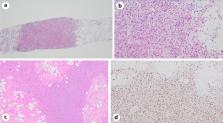
          Solitary fibrous tumor (SFT) is an uncommon tumor of mesenchymal origin, which can arise at any anatomic location and can exhibit versatile histological features and a clinical course ranging from benign to frankly malignant. Lipomatous (fat-forming) SFT is a morphological variant of SFT characterized by an adipose tissue component. Breast SFT is an extremely rare clinical entity, and the literature review yielded only 28 previously reported cases. However, lipomatous (fat-forming) SFT is much less common than conventional tumors and, to our knowledge, it has never been reported in the breast. We describe a case of a 54-year-old postmenopausal woman who presented with a palpable mass on her right breast. No other associated features such as nipple discharge, skin changes, or axillary lymphadenopathy were present. The clinical differential diagnosis included fibroadenoma, phyllodes tumor, and mammary hamartoma. A ultrasound scan examination demonstrated a large, oval, well-circumscribed lesion with indeterminate features, but suspicious of malignancy. However, a needle core biopsy was performed and histological examination with ancillary immunohistochemical staining confirmed the diagnosis of SFT, a lipomatous variant. The lesion was excised with clear margins and histological examination confirmed SFT with low-risk features and follow-up was planned. Careful histological evaluation with diffuse and strong nuclear expression of STAT6 helped to distinguish lipomatous SFTs from other mimics. Here, we describe the first case of a lipomatous variant of a SFT involving the breast.

          Related collections

          Most cited references15

          • Record: found
          • Abstract: found
          • Article: not found

          Nuclear expression of STAT6 distinguishes solitary fibrous tumor from histologic mimics.

          Solitary fibrous tumor (SFT) is composed of spindled to ovoid cells in a patternless architecture with prominent stromal collagen and hemangiopericytoma-like vessels. Some tumors show hypercellularity, nuclear atypia, and significant mitotic activity; the latter feature in particular often portends an aggressive clinical course. SFT can sometimes be difficult to distinguish from other benign mesenchymal tumors and sarcomas. The most characteristic (albeit nonspecific) immunohistochemical finding in SFT is CD34 expression. A NAB2-STAT6 gene fusion, resulting in a chimeric protein in which a repressor domain of NGFI-A binding protein 2 (EGR1 binding protein 2) (NAB2) is replaced with a carboxy-terminal transactivation domain from signal transducer and activator of transcription 6, interleukin-4 induced (STAT6), was recently identified as a consistent finding in SFT. However, as these genes are located in close proximity on 12q13, this fusion can only rarely be detected by conventional chromosomal banding or fluorescence in situ hybridization analysis. Nuclear expression of the carboxy terminal part of STAT6 is a consistent finding in SFT of the meninges (so-called 'meningeal hemangiopericytoma'). We investigated STAT6 expression by immunohistochemistry in SFTs and other soft tissue tumors arising outside the central nervous system to validate the diagnostic utility of this novel marker. Whole-tissue sections of 231 tumors were evaluated, including 60 cases of SFT as well as other benign and malignant mesenchymal neoplasms and sarcomatoid mesotheliomas. Fifty-nine of 60 SFT cases (98%) showed nuclear expression of STAT6, which was usually diffuse and intense. All other tumor types were negative for STAT6, except for three dedifferentiated liposarcomas and one deep fibrous histiocytoma, which showed weak staining. In conclusion, STAT6 is a highly sensitive and almost perfectly specific immunohistochemical marker for SFT and can be helpful to distinguish this tumor type from histologic mimics.
            Bookmark
            • Record: found
            • Abstract: found
            • Article: not found

            STAT6 immunohistochemistry is helpful in the diagnosis of solitary fibrous tumors.

            Solitary fibrous tumor (SFT) is an uncommon fibroblastic neoplasm. Although histologic characteristics and frequent CD34 expression allow for an accurate diagnosis in the majority of SFT cases, a wide histologic spectrum and an occasional unexpected immunophenotype may pose diagnostic challenges. Molecular analyses have discovered that almost all SFTs harbor an NAB2-STAT6 fusion gene, which is considered specific to this tumor type. Recent studies have suggested that STAT6 immunohistochemistry is a reliable surrogate for detection of the fusion gene. Our aim was to validate these findings by examining a large number of SFT cases and a broad array of 30 different types of non-SFT tumors. A total of 49 SFTs with a range of histologic characteristics and 159 benign or malignant tumors that can mimic SFTs were retrieved and stained for STAT6. All 49 SFTs (100%) showed STAT6 expression that was restricted in the nucleus, mostly in a diffuse and strong manner, irrespective of the tumor sites and histologic patterns. The staining was uniform in most cases but was heterogenous in about 20% of the cases in which zonal staining attenuation was observed, likely reflecting variability in fixation or tissue ischemia. In contrast, only 4 non-SFT tumors (2.5%) exhibited weak nuclear STAT6 expression, whereas the remaining 155 cases showed no staining or often weak reactivity in both the cytoplasm and the nucleus. Therefore, nuclear STAT6 immunoreactivity is a highly sensitive and specific marker of SFTs and can be helpful when diagnosis is inconclusive by conventional methods.
              Bookmark
              • Record: found
              • Abstract: not found
              • Article: not found

              Risk assessment in solitary fibrous tumors: validation and refinement of a risk stratification model

                Bookmark

                Author and article information

                Journal
                Case Rep Oncol
                Case Rep Oncol
                CRO
                Case Reports in Oncology
                S. Karger AG (Allschwilerstrasse 10, P.O. Box · Postfach · Case postale, CH–4009, Basel, Switzerland · Schweiz · Suisse, Phone: +41 61 306 11 11, Fax: +41 61 306 12 34, karger@karger.com )
                1662-6575
                Jan-Apr 2022
                28 April 2022
                28 April 2022
                : 15
                : 1
                : 455-461
                Affiliations
                [1] aPathology Department, Sheikh Shakhbout Medical City, Abu Dhabi, United Arab Emirates
                [2] bGeneral Surgery Department, Sheikh Shakhbout Medical City, Abu Dhabi, United Arab Emirates
                Author notes
                *Najla Saleh Ben Ghashir, nsghashir@ 123456ssmc.ae
                Article
                cro-0015-0455
                10.1159/000524364
                9149475
                35702562
                da481dad-164f-4017-8ea8-b9e0ac12f0b0
                Copyright © 2022 by S. Karger AG, Basel

                This article is licensed under the Creative Commons Attribution-NonCommercial-4.0 International License (CC BY-NC) (http://www.karger.com/Services/OpenAccessLicense). Usage and distribution for commercial purposes requires written permission.

                History
                : 15 March 2022
                : 20 March 2022
                : 2022
                Page count
                Figures: 2, References: 15, Pages: 7
                Categories
                Case Report

                Oncology & Radiotherapy
                breast,lipomatous,solitary fibrous tumor
                Oncology & Radiotherapy
                breast, lipomatous, solitary fibrous tumor

                Comments

                Comment on this article