14
views
0
recommends
+1 Recommend
0 collections
    0
    shares
      • Record: found
      • Abstract: found
      • Article: found
      Is Open Access

      Dual Roles for Membrane Association of Drosophila Axin in Wnt Signaling

      research-article
      , , , *
      PLoS Genetics
      Public Library of Science

      Read this article at

      Bookmark
          There is no author summary for this article yet. Authors can add summaries to their articles on ScienceOpen to make them more accessible to a non-specialist audience.

          Abstract

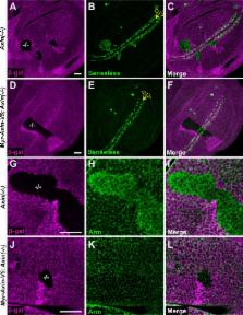
          Deregulation of the Wnt signal transduction pathway underlies numerous congenital disorders and cancers. Axin, a concentration-limiting scaffold protein, facilitates assembly of a “destruction complex” that prevents signaling in the unstimulated state and a plasma membrane-associated “signalosome” that activates signaling following Wnt stimulation. In the classical model, Axin is cytoplasmic under basal conditions, but relocates to the cell membrane after Wnt exposure; however, due to the very low levels of endogenous Axin, this model is based largely on examination of Axin at supraphysiological levels. Here, we analyze the subcellular distribution of endogenous Drosophila Axin in vivo and find that a pool of Axin localizes to cell membrane proximal puncta even in the absence of Wnt stimulation. Axin localization in these puncta is dependent on the destruction complex component Adenomatous polyposis coli (Apc). In the unstimulated state, the membrane association of Axin increases its Tankyrase-dependent ADP-ribosylation and consequent proteasomal degradation to control its basal levels. Furthermore, Wnt stimulation does not result in a bulk redistribution of Axin from cytoplasmic to membrane pools, but causes an initial increase of Axin in both of these pools, with concomitant changes in two post-translational modifications, followed by Axin proteolysis hours later. Finally, the ADP-ribosylated Axin that increases rapidly following Wnt stimulation is membrane associated. We conclude that even in the unstimulated state, a pool of Axin forms membrane-proximal puncta that are dependent on Apc, and that membrane association regulates both Axin levels and Axin’s role in the rapid activation of signaling that follows Wnt exposure.

          Author Summary

          Axin is a scaffold protein with essential roles in Wnt signal transduction. In the classical model, the transition from the unstimulated to stimulated state is thought to be achieved by recruitment of Axin from cytosol to plasma membrane. We find that a pool of endogenous Drosophila Axin is localized in puncta juxtaposed with the cell membrane even under basal conditions and is targeted for degradation by the ADP-ribose polymerase Tankyrase. Wnt stimulation initially results in increased Axin levels in both the cytosolic and membrane pools, which may enhance the robust activation of signaling.

          Related collections

          Most cited references63

          • Record: found
          • Abstract: found
          • Article: not found

          Tankyrase inhibition stabilizes axin and antagonizes Wnt signalling.

          The stability of the Wnt pathway transcription factor beta-catenin is tightly regulated by the multi-subunit destruction complex. Deregulated Wnt pathway activity has been implicated in many cancers, making this pathway an attractive target for anticancer therapies. However, the development of targeted Wnt pathway inhibitors has been hampered by the limited number of pathway components that are amenable to small molecule inhibition. Here, we used a chemical genetic screen to identify a small molecule, XAV939, which selectively inhibits beta-catenin-mediated transcription. XAV939 stimulates beta-catenin degradation by stabilizing axin, the concentration-limiting component of the destruction complex. Using a quantitative chemical proteomic approach, we discovered that XAV939 stabilizes axin by inhibiting the poly-ADP-ribosylating enzymes tankyrase 1 and tankyrase 2. Both tankyrase isoforms interact with a highly conserved domain of axin and stimulate its degradation through the ubiquitin-proteasome pathway. Thus, our study provides new mechanistic insights into the regulation of axin protein homeostasis and presents new avenues for targeted Wnt pathway therapies.
            Bookmark
            • Record: found
            • Abstract: found
            • Article: not found

            Analysis of genetic mosaics in developing and adult Drosophila tissues.

            T Xu, G Rubin (1993)
            We have constructed a series of strains to facilitate the generation and analysis of clones of genetically distinct cells in developing and adult tissues of Drosophila. Each of these strains carries an FRT element, the target for the yeast FLP recombinase, near the base of a major chromosome arm, as well as a gratuitous cell-autonomous marker. Novel markers that carry epitope tags and that are localized to either the cell nucleus or cell membrane have been generated. As a demonstration of how these strains can be used to study a particular gene, we have analyzed the developmental role of the Drosophila EGF receptor homolog. Moreover, we have shown that these strains can be utilized to identify new mutations in mosaic animals in an efficient and unbiased way, thereby providing an unprecedented opportunity to perform systematic genetic screens for mutations affecting many biological processes.
              Bookmark
              • Record: found
              • Abstract: found
              • Article: not found

              Wnt induces LRP6 signalosomes and promotes dishevelled-dependent LRP6 phosphorylation.

              Multiple signaling pathways, including Wnt signaling, participate in animal development, stem cell biology, and human cancer. Although many components of the Wnt pathway have been identified, unresolved questions remain as to the mechanism by which Wnt binding to its receptors Frizzled and Low-density lipoprotein receptor-related protein 6 (LRP6) triggers downstream signaling events. With live imaging of vertebrate cells, we show that Wnt treatment quickly induces plasma membrane-associated LRP6 aggregates. LRP6 aggregates are phosphorylated and can be detergent-solubilized as ribosome-sized multiprotein complexes. Phospho-LRP6 aggregates contain Wnt-pathway components but no common vesicular traffic markers except caveolin. The scaffold protein Dishevelled (Dvl) is required for LRP6 phosphorylation and aggregation. We propose that Wnts induce coclustering of receptors and Dvl in LRP6-signalosomes, which in turn triggers LRP6 phosphorylation to promote Axin recruitment and beta-catenin stabilization.
                Bookmark

                Author and article information

                Contributors
                Role: Editor
                Journal
                PLoS Genet
                PLoS Genet
                plos
                plosgen
                PLoS Genetics
                Public Library of Science (San Francisco, CA USA )
                1553-7390
                1553-7404
                13 December 2016
                December 2016
                : 12
                : 12
                : e1006494
                Affiliations
                [001]Department of Molecular and Systems Biology and the Norris Cotton Cancer Center, Geisel School of Medicine at Dartmouth College, Hanover, NH, United States of America
                University of Michigan, UNITED STATES
                Author notes

                The authors have declared that no competing interests exist.

                • Conceptualization: ZW OTB YA.

                • Investigation: ZW OTB EY YA.

                • Writing – original draft: ZW OTB YA.

                Article
                PGENETICS-D-16-00607
                10.1371/journal.pgen.1006494
                5154497
                27959917
                d9aa1254-8228-473b-a668-8d1ebfd8d67b
                © 2016 Wang et al

                This is an open access article distributed under the terms of the Creative Commons Attribution License, which permits unrestricted use, distribution, and reproduction in any medium, provided the original author and source are credited.

                History
                : 16 March 2016
                : 17 November 2016
                Page count
                Figures: 8, Tables: 0, Pages: 26
                Funding
                Funded by: funder-id http://dx.doi.org/10.13039/100000002, National Institutes of Health;
                Award ID: RO1CA105038
                Award Recipient :
                Funded by: funder-id http://dx.doi.org/10.13039/100000002, National Institutes of Health;
                Award ID: P40OD018537
                Award Recipient :
                Funded by: Emerald Foundation
                Award Recipient :
                Funded by: the Norris Cotton Cancer Center
                Award Recipient :
                Funded by: funder-id http://dx.doi.org/10.13039/100000001, National Science Foundation;
                Award ID: DBI-1039423
                This work was funded by grants from the NIH (RO1CA105038 to YA and P40OD018537 to the BDSC), Emerald Foundation, the Norris Cotton Cancer Center and the National Science Foundation (DBI-1039423 for purchase of a Nikon A1RSi confocal microscope). The funders had no role in study design, data collection and analysis, decision to publish, or preparation of the manuscript.
                Categories
                Research Article
                Biology and Life Sciences
                Cell Biology
                Cellular Structures and Organelles
                Cell Membranes
                Biology and Life Sciences
                Molecular Biology
                Molecular Biology Techniques
                Cloning
                Research and Analysis Methods
                Molecular Biology Techniques
                Cloning
                Research and Analysis Methods
                Model Organisms
                Animal Models
                Drosophila Melanogaster
                Biology and Life Sciences
                Organisms
                Animals
                Invertebrates
                Arthropoda
                Insects
                Drosophila
                Drosophila Melanogaster
                Physical sciences
                Chemistry
                Chemical reactions
                ADP-ribosylation
                Biology and Life Sciences
                Cell Biology
                Cellular Structures and Organelles
                Cytoplasm
                Research and Analysis Methods
                Specimen Preparation and Treatment
                Staining
                Membrane Staining
                Biology and Life Sciences
                Molecular Biology
                Molecular Biology Techniques
                Molecular Probe Techniques
                Immunoblotting
                Research and Analysis Methods
                Molecular Biology Techniques
                Molecular Probe Techniques
                Immunoblotting
                Biology and Life Sciences
                Developmental Biology
                Embryology
                Embryos
                Custom metadata
                All relevant data are within the paper and its Supporting Information files.

                Genetics
                Genetics

                Comments

                Comment on this article