2
views
0
recommends
+1 Recommend
0 collections
    0
    shares
      • Record: found
      • Abstract: found
      • Article: found
      Is Open Access

      Applying quantitative spatial phenotypes analysis to the investigation of peltate glandular trichomes development pattern in Perilla frutescens

      research-article

      Read this article at

      Bookmark
          There is no author summary for this article yet. Authors can add summaries to their articles on ScienceOpen to make them more accessible to a non-specialist audience.

          Abstract

          Background

          Glandular trichomes, often referred to as “phytochemical factories”, plays a crucial role in plant growth and metabolism. As the site for secretion and storage, the development of glandular trichomes is related to the dynamic biosynthesis of specialised metabolites. The study aims to explore the relationship between spatial phenotype and dynamic metabolism of glandular trichomes, and establish a novel approach for the exploration and study of the regulatory mechanism governing the development of glandular trichomes.

          Results

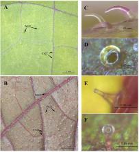
          In this study, we proposed a technical route based on the relative deviation value to distinguish the peltate glandular trichomes (PGTs) from the background tissues and extract their spatial phenotype. By defining glandular trichome developmental stages based on the leaf vein growth axis, we found that young PGTs were densely distributed near the proximal end of growth axis of the leaf veins, where perillaketone, a primary metabolite of PGTs, is predominantly accumulated. Conversely, mature PGTs are typically found near the distal end of the mid-vein growth axis and the lateral end of the secondary vein growth axis, where the accumulation rate of isoegomaketone and egomaketone exceeds that of perillaketone in PGTs. We further identified spatial phenotypic parameters, L sum and d, as independent variables to construct a linear regression model that illustrates the relationship between the spatial phenotypes and metabolite content of PGTs, including perillaketone (R 2 = 0.698), egomaketone (R 2 = 0.593), isoegomaketone (R 2 = 0.662) and the sum of the amount (R 2 = 0.773).

          Conclusions

          This model proved that the development of PGTs was correlated with the growth of the entire leaf, and the development stage of PGTs can be identifined by spatial phenotypes based on the leaf veins. In conclusion, the findings of this study enhance our understanding of correlation between spatial phenotype and development of glandular trichomes and offer a new approach to explore and study the regulatory mechanism of glandular trichome development.

          Supplementary Information

          The online version contains supplementary material available at 10.1186/s13007-023-01072-4.

          Related collections

          Most cited references60

          • Record: found
          • Abstract: found
          • Article: found
          Is Open Access

          Combined single-cell and spatial transcriptomics reveals the molecular, cellular and spatial bone marrow niche organization

          The bone marrow (BM) constitutes the primary site for life-long blood production and skeletal regeneration. However, its cellular composition and the spatial organization into distinct ‘niches’ remains controversial. Here, we combine single-cell and spatially resolved transcriptomics to systematically map the molecular and cellular composition of the endosteal, sinusoidal, and arteriolar BM niches. This allowed us to transcriptionally profile all major BM resident cell types, determine their localization, and clarify the cellular and spatial sources of key growth factors and cytokines. Our data demonstrate that previously unrecognized Cxcl12-abundant reticular (CAR) cell subsets (i.e. Adipo- and Osteo- CAR cells) differentially localize to sinusoidal or arteriolar surfaces, locally act as ‘professional cytokine secreting cells’, and thereby establish distinct peri-vascular micro-niches. Importantly, we also demonstrate that the 3-dimensional organization of the BM can be accurately inferred from single-cell gene expression data using the newly developed RNA-Magnet algorithm. Together, our study reveals the cellular and spatial organization of BM niches, and offers a novel strategy to dissect the complex organization of whole organs in a systematic manner.
            Bookmark
            • Record: found
            • Abstract: found
            • Article: not found
            Is Open Access

            Spatiotemporal transcriptomic atlas of mouse organogenesis using DNA nanoball-patterned arrays

            Spatially resolved transcriptomic technologies are promising tools to study complex biological processes such as mammalian embryogenesis. However, the imbalance between resolution, gene capture, and field of view of current methodologies precludes their systematic application to analyze relatively large and three-dimensional mid- and late-gestation embryos. Here, we combined DNA nanoball (DNB)-patterned arrays and in situ RNA capture to create spatial enhanced resolution omics-sequencing (Stereo-seq). We applied Stereo-seq to generate the mouse organogenesis spatiotemporal transcriptomic atlas (MOSTA), which maps with single-cell resolution and high sensitivity the kinetics and directionality of transcriptional variation during mouse organogenesis. We used this information to gain insight into the molecular basis of spatial cell heterogeneity and cell fate specification in developing tissues such as the dorsal midbrain. Our panoramic atlas will facilitate in-depth investigation of longstanding questions concerning normal and abnormal mammalian development.
              Bookmark
              • Record: found
              • Abstract: found
              • Article: not found

              Exit from proliferation during leaf development in Arabidopsis thaliana: a not-so-gradual process.

              Early leaf growth is sustained by cell proliferation and subsequent cell expansion that initiates at the leaf tip and proceeds in a basipetal direction. Using detailed kinematic and gene expression studies to map these stages during early development of the third leaf of Arabidopsis thaliana, we showed that the cell-cycle arrest front did not progress gradually down the leaf, but rather was established and abolished abruptly. Interestingly, leaf greening and stomatal patterning followed a similar basipetal pattern, but proliferative pavement cell and formative meristemoid divisions were uncoordinated in respect to onset and persistence. Genes differentially expressed during the transition from cell proliferation to expansion were enriched in genes involved in cell cycle, photosynthesis, and chloroplast retrograde signaling. Proliferating primordia treated with norflurazon, a chemical inhibitor of retrograde signaling, showed inhibited onset of cell expansion. Hence, differentiation of the photosynthetic machinery is important for regulating the exit from proliferation. Copyright © 2012 Elsevier Inc. All rights reserved.
                Bookmark

                Author and article information

                Contributors
                jyjiangzheng@163.com
                20203098@njucm.edu.cn
                20200622@njucm.edu.cn
                1787489884@qq.com
                yuewei@njucm.edu.cn
                qucheng@njucm.edu.cn
                wuqn@njucm.edu.cn
                Journal
                Plant Methods
                Plant Methods
                Plant Methods
                BioMed Central (London )
                1746-4811
                25 August 2023
                25 August 2023
                2023
                : 19
                : 88
                Affiliations
                [1 ]GRID grid.410745.3, ISNI 0000 0004 1765 1045, College of Pharmacy, , Nanjing University of Chinese Medicine, ; Nanjing, 210023 China
                [2 ]Collaborative Innovation Center of Chinese Medicinal Resources Industrialization, Nanjing, 210023 China
                [3 ]National and Local Collaborative Engineering Center of Chinese Medicinal Resources Industrialization and Formulae Innovative Medicine, Nanjing, 210023 China
                Article
                1072
                10.1186/s13007-023-01072-4
                10464211
                37626389
                d958ead3-cb51-44ea-ac82-8e08249638aa
                © BioMed Central Ltd., part of Springer Nature 2023

                Open Access This article is licensed under a Creative Commons Attribution 4.0 International License, which permits use, sharing, adaptation, distribution and reproduction in any medium or format, as long as you give appropriate credit to the original author(s) and the source, provide a link to the Creative Commons licence, and indicate if changes were made. The images or other third party material in this article are included in the article's Creative Commons licence, unless indicated otherwise in a credit line to the material. If material is not included in the article's Creative Commons licence and your intended use is not permitted by statutory regulation or exceeds the permitted use, you will need to obtain permission directly from the copyright holder. To view a copy of this licence, visit http://creativecommons.org/licenses/by/4.0/. The Creative Commons Public Domain Dedication waiver ( http://creativecommons.org/publicdomain/zero/1.0/) applies to the data made available in this article, unless otherwise stated in a credit line to the data.

                History
                : 24 December 2022
                : 16 August 2023
                Funding
                Funded by: The research of ecological planting and quality assurance of Jiangsu Dao-di herbs
                Award ID: 2022
                Funded by: Chinese traditional medicine resource security enhancement
                Award ID: 2023
                Categories
                Methodology
                Custom metadata
                © BioMed Central Ltd., part of Springer Nature 2023

                Plant science & Botany
                growth axis,linear regression equation,leaf veins,peltate glandular trichomes,perilla frutescens,spatial phenotype

                Comments

                Comment on this article