0
views
0
recommends
+1 Recommend
0 collections
    0
    shares
      • Record: found
      • Abstract: found
      • Article: found
      Is Open Access

      Periarticular muscle status affects in vivo tibio-femoral joint loads after total knee arthroplasty

      research-article

      Read this article at

      Bookmark
          There is no author summary for this article yet. Authors can add summaries to their articles on ScienceOpen to make them more accessible to a non-specialist audience.

          Abstract

          Background: Total knee arthroplasty (TKA) is a highly effective treatment for severe knee osteoarthritis that is increasingly performed in younger, more active patients. As postoperative muscular impairments may negatively affect surgical outcomes and implant longevity, functional muscle recovery gains increasing importance in meeting future patient demands. This study aimed to assess the status of periarticular muscles in the long-term follow-up after TKA and to evaluate its impact on in vivo tibio-femoral joint loads.

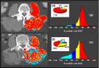
          Methods: A case series was created, with eight patients with knee osteoarthritis. All subjects received an instrumented knee implant in unilateral TKA. Native computed tomography scans, acquired pre and postoperatively, were used to evaluate distal muscle volumes and fatty infiltration. In vivo tibio-femoral joint loads were measured telemetrically during standing, walking, stair climbing and chair rising and were correlated to muscle status.

          Results: Postoperatively a reduction in fatty infiltration across all periarticular muscles was pronounced. High average peak loads acted in the tibio-femoral joint ranging from 264% during stand-to-sit activities up to 341% body weight (BW) during stair descent. Fatty infiltration of the m. quadriceps femoris and hamstrings were associated with increased tibio-femoral joint contact forces during walking (r = 0.542; 0.412 and 0.766).

          Conclusion: The findings suggest that a fatty infiltration of periarticular muscles may lead to increased tibio-femoral joint contact forces. However, we only observed weak correlations between these parameters. Improvements in functional mobility and the restoration of a pain-free joint likely explain the observed postoperative reductions in fatty infiltration. Perioperative rehabilitation approaches targeting residual impairments in muscle quality could, contribute to reduced tibio-femoral joint loads and improved long-term outcomes of TKA. However, it has to be pointed out that the study included a small number of patients, which may limit its validity.

          Related collections

          Most cited references51

          • Record: found
          • Abstract: found
          • Article: not found

          Loading of the knee joint during activities of daily living measured in vivo in five subjects.

          Detailed knowledge about loading of the knee joint is essential for preclinical testing of implants, validation of musculoskeletal models and biomechanical understanding of the knee joint. The contact forces and moments acting on the tibial component were therefore measured in 5 subjects in vivo by an instrumented knee implant during various activities of daily living. Average peak resultant forces, in percent of body weight, were highest during stair descending (346% BW), followed by stair ascending (316% BW), level walking (261% BW), one legged stance (259% BW), knee bending (253% BW), standing up (246% BW), sitting down (225% BW) and two legged stance (107% BW). Peak shear forces were about 10-20 times smaller than the axial force. Resultant forces acted almost vertically on the tibial plateau even during high flexion. Highest moments acted in the frontal plane with a typical peak to peak range -2.91% BWm (adduction moment) to 1.61% BWm (abduction moment) throughout all activities. Peak flexion/extension moments ranged between -0.44% BWm (extension moment) and 3.16% BWm (flexion moment). Peak external/internal torques lay between -1.1% BWm (internal torque) and 0.53% BWm (external torque). The knee joint is highly loaded during daily life. In general, resultant contact forces during dynamic activities were lower than the ones predicted by many mathematical models, but lay in a similar range as measured in vivo by others. Some of the observed load components were much higher than those currently applied when testing knee implants. Copyright 2010 Elsevier Ltd. All rights reserved.
            Bookmark
            • Record: found
            • Abstract: found
            • Article: found
            Is Open Access

            The effect of patient age at intervention on risk of implant revision after total replacement of the hip or knee: a population-based cohort study

            Summary Background Total joint replacements for end-stage osteoarthritis of the hip and knee are cost-effective and demonstrate significant clinical improvement. However, robust population based lifetime-risk data for implant revision are not available to aid patient decision making, which is a particular problem in young patient groups deciding on best-timing for surgery. Methods We did implant survival analysis on all patients within the Clinical Practice Research Datalink who had undergone total hip replacement or total knee replacement. These data were adjusted for all-cause mortality with data from the Office for National Statistics and used to generate lifetime risks of revision surgery based on increasing age at the time of primary surgery. Findings We identified 63 158 patients who had undergone total hip replacement and 54 276 who had total knee replacement between Jan 1, 1991, and Aug 10, 2011, and followed up these patients to a maximum of 20 years. For total hip replacement, 10-year implant survival rate was 95·6% (95% CI 95·3–95·9) and 20-year rate was 85·0% (83·2–86·6). For total knee replacement, 10-year implant survival rate was 96·1% (95·8–96·4), and 20-year implant survival rate was 89·7% (87·5–91·5). The lifetime risk of requiring revision surgery in patients who had total hip replacement or total knee replacement over the age of 70 years was about 5% with no difference between sexes. For those who had surgery younger than 70 years, however, the lifetime risk of revision increased for younger patients, up to 35% (95% CI 30·9–39·1) for men in their early 50s, with large differences seen between male and female patients (15% lower for women in same age group). The median time to revision for patients who had surgery younger than age 60 was 4·4 years. Interpretation Our study used novel methodology to investigate and offer new insight into the importance of young age and risk of revision after total hip or knee replacement. Our evidence challenges the increasing trend for more total hip replacements and total knee replacements to be done in the younger patient group, and these data should be offered to patients as part of the shared decision making process. Funding Oxford Musculoskeletal Biomedical Research Unit, National Institute for Health Research.
              Bookmark
              • Record: found
              • Abstract: found
              • Article: found
              Is Open Access

              Measurement of skeletal muscle radiation attenuation and basis of its biological variation

              Skeletal muscle contains intramyocellular lipid droplets within the cytoplasm of myocytes as well as intermuscular adipocytes. These depots exhibit physiological and pathological variation which has been revealed with the advent of diagnostic imaging approaches: magnetic resonance (MR) imaging, MR spectroscopy and computed tomography (CT). CT uses computer-processed X-rays and is now being applied in muscle physiology research. The purpose of this review is to present CT methodologies and summarize factors that influence muscle radiation attenuation, a parameter which is inversely related to muscle fat content. Pre-defined radiation attenuation ranges are used to demarcate intermuscular adipose tissue [from −190 to −30 Hounsfield units (HU)] and muscle (−29 HU to +150 HU). Within the latter range, the mean muscle radiation attenuation [muscle (radio) density] is reported. Inconsistent criteria for the upper and lower HU cut-offs used to characterize muscle attenuation limit comparisons between investigations. This area of research would benefit from standardized criteria for reporting muscle attenuation. Available evidence suggests that muscle attenuation is plastic with physiological variation induced by the process of ageing, as well as by aerobic training, which probably reflects accumulation of lipids to fuel aerobic work. Pathological variation in muscle attenuation reflects excess fat deposition in the tissue and is observed in people with obesity, diabetes type II, myositis, osteoarthritis, spinal stenosis and cancer. A poor prognosis and different types of morbidity are predicted by the presence of reduced mean muscle attenuation values in patients with these conditions; however, the biological features of muscle with these characteristics require further investigation.
                Bookmark

                Author and article information

                Contributors
                Journal
                Front Bioeng Biotechnol
                Front Bioeng Biotechnol
                Front. Bioeng. Biotechnol.
                Frontiers in Bioengineering and Biotechnology
                Frontiers Media S.A.
                2296-4185
                22 March 2023
                2023
                : 11
                : 1075357
                Affiliations
                [1] 1 Berlin Institute of Health at Charité—Universitätsmedizin Berlin , Center for Musculoskeletal Biomechanics and Regeneration (Julius Wolff Institute) , Berlin, Germany
                [2] 2 Berlin Institute of Health at Charité—Universitätsmedizin Berlin , Berlin Institute of Health Institute for Regenerative Therapies , Berlin, Germany
                [3] 3 Center for Musculoskeletal Surgery , Charité—Universitätsmedizin Berlin , Berlin, Germany
                [4] 4 Department of Radiology , Charité University Medicine Berlin , Berlin, Germany
                Author notes

                Edited by: Ajay Seth, Delft University of Technology, Netherlands

                Reviewed by: Hans Kainz, University of Vienna, Austria

                Yaodong Gu, Ningbo University, China

                *Correspondence: Philipp Damm, philipp.damm@ 123456bih-charite.de

                This article was submitted to Biomechanics, a section of the journal Frontiers in Bioengineering and Biotechnology

                Article
                1075357
                10.3389/fbioe.2023.1075357
                10073542
                37034264
                d837317c-6661-4c75-89fe-800500d0e7a6
                Copyright © 2023 Winkler, Bell, Bender, Trepczynski, Duda, Baur and Damm.

                This is an open-access article distributed under the terms of the Creative Commons Attribution License (CC BY). The use, distribution or reproduction in other forums is permitted, provided the original author(s) and the copyright owner(s) are credited and that the original publication in this journal is cited, in accordance with accepted academic practice. No use, distribution or reproduction is permitted which does not comply with these terms.

                History
                : 20 October 2022
                : 09 March 2023
                Funding
                Funded by: Deutsche Forschungsgemeinschaft , doi 10.13039/501100001659;
                Funded by: Bundesministerium für Bildung, Wissenschaft und Forschung , doi 10.13039/501100013699;
                This study was supported by the German Research Foundation (DFG) (TR 1657/1-1, DA 1786/5-1, SFB 1444 (427826188)), the Federal Ministry of Education and Research (workHEALTH (01EC 1905D) and the OrthoLoadClub. The funding bodies had no role in the study design, collection and analysis of data, writing of the manuscript and decision to publish.
                Categories
                Bioengineering and Biotechnology
                Original Research

                total knee arthroplasty (replacement),instrumented knee implants,periarticular muscle status, in vivo tibio-femoral joint loads, in vivo measurement

                Comments

                Comment on this article