21
views
0
recommends
+1 Recommend
0 collections
    0
    shares
      • Record: found
      • Abstract: found
      • Article: found
      Is Open Access

      Hybrid fracture fixation systems developed for orthopaedic applications: A general review

      review-article

      Read this article at

      Bookmark
          There is no author summary for this article yet. Authors can add summaries to their articles on ScienceOpen to make them more accessible to a non-specialist audience.

          Abstract

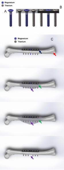
          Orthopaedic implants are applied daily in our orthopaedic clinics for treatment of musculoskeletal injuries, especially for bone fracture fixation. To realise the multiple functions of orthopaedic implants, hybrid system that contains several different materials or parts have also been designed for application, such as prosthesis for total hip arthroplasty. Fixation of osteoporotic fracture is challenging as the current metal implants made of stainless steel or titanium that are rather rigid and bioinert, which are not favourable for enhancing fracture healing and subsequent remodelling. Magnesium (Mg) and its alloys are reported to possess good biocompatibility, biodegradability and osteopromotive effects during its in vivo degradation and now tested as a new generation of degradable metallic biomaterials. Several recent clinical studies reported the Mg-based screws for bone fixation, although the history of testing Mg as fixation implant was documented more than 100 years ago. Truthfully, Mg has its limitations as fixation implant, especially when applied at load-bearing sites because of rather rapid degradation. Currently developed Mg-based implants have only been designed for application at less or non–loading-bearing skeletal site(s). Therefore, after years research and development, the authors propose an innovative hybrid fixation system with parts composed of Mg and titanium or stainless steel to maximise the biological benefits of Mg; titanium or stainless steel in this hybrid system can provide enough mechanical support for fractures at load-bearing site(s) while Mg promotes the fracture healing through novel mechanisms during its degradation, especially in patients with osteoporosis and other metabolic disorders that are unfavourable conditions for fracture healing. This hybrid fixation strategy is designed to effectively enhance the osteoporotic fracture healing and may potentially also reduce the refracture rate.

          The translational potential of this article: This article systemically reviewed the combination utility of different metallic implants in orthopaedic applications. It will do great contribution to the further development of internal orthopaedic implants for fracture fixation. Meanwhile, it also introduced a titanium–magnesium hybrid fixation system as an alternative fixation strategy, especially for osteoporotic patients.

          Related collections

          Most cited references105

          • Record: found
          • Abstract: found
          • Article: not found

          The history of biodegradable magnesium implants: a review.

          Today, more than 200years after the first production of metallic magnesium by Sir Humphry Davy in 1808, biodegradable magnesium-based metal implants are currently breaking the paradigm in biomaterial science to develop only highly corrosion resistant metals. This groundbreaking approach to temporary metallic implants is one of the latest developments in biomaterials science that is being rediscovered. It is a challenging topic, and several secrets still remain that might revolutionize various biomedical implants currently in clinical use. Magnesium alloys were investigated as implant materials long ago. A very early clinical report was given in 1878 by the physician Edward C. Huse. He used magnesium wires as ligature for bleeding vessels. Magnesium alloys for clinical use were explored during the last two centuries mainly by surgeons with various clinical backgrounds, such as cardiovascular, musculoskeletal and general surgery. Nearly all patients benefited from the treatment with magnesium implants. Although most patients experienced subcutaneous gas cavities caused by rapid implant corrosion, most patients had no pain and almost no infections were observed during the postoperative follow-up. This review critically summarizes the in vitro and in vivo knowledge and experience that has been reported on the use of magnesium and its alloys to advance the field of biodegradable metals. Copyright (c) 2010 Acta Materialia Inc. Published by Elsevier Ltd. All rights reserved.
            Bookmark
            • Record: found
            • Abstract: found
            • Article: not found

            In vitro and in vivo corrosion measurements of magnesium alloys.

            The in vivo corrosion of magnesium alloys might provide a new mechanism which would allow degradable metal implants to be applied in musculo-skeletal surgery. This would particularly be true if magnesium alloys with controlled in vivo corrosion rates could be developed. Since the magnesium corrosion process depends on its corrosive environment, the corrosion rates of magnesium alloys under standard in vitro environmental conditions were compared to corrosion rates in an in vivo animal model. Two gravity-cast magnesium alloys (AZ91D, LAE442) were used in these investigations. Standardized immersion and electrochemical tests according to ASTM norms were performed. The in vivo corrosion tests were carried out by intramedullar implantation of sample rods of the magnesium alloys in guinea pig femura. The reduction in implant volume was determined by synchrotron-radiation-based microtomography. We found that in vivo corrosion was about four orders of magnitude lower than in vitro corrosion of the tested alloys. Furthermore, the tendency of the corrosion rates obtained from in vitro corrosion tests were in the opposite direction as those obtained from the in vivo study. The results of this study suggest, that the conclusions drawn from current ASTM standard in vitro corrosion tests cannot be used to predict in vivo corrosion rates of magnesium alloys.
              Bookmark
              • Record: found
              • Abstract: found
              • Article: not found

              The cell and molecular biology of fracture healing.

              Fracture healing is a complex physiologic process that involves the coordinated participation of several cell types. By using a reproducible model of experimental fracture healing in the rat, it is possible to elucidate the integrated cellular responses that signal the pathways and the role of the extracellular matrix components in orchestrating the events of fracture healing. Histologic characterization of fracture healing shows that intramembranous ossification occurs under the periosteum within a few days after an injury. Events of endochondral ossification occur adjacent to the fracture site and span a period of up to 28 days. Remodeling of the woven bone formed by intramembranous and endochondral ossification proceeds for several weeks. Spatial and temporal expression of genes for major collagens (Types I and II), minor fibrillar collagens (Types IV and XI), and several extracellular matrix components (osteocalcin, osteonectin, osteopontin, fibronectin and CD44) are detected by in situ hybridization. Immunohistochemical studies show that expression of proliferating cell nuclear antigen is both time and space dependent and differentially expressed in the callus tissues formed by the intramembranous and endochondral processes. Chondrocytes involved in endochondral ossification undergo apoptosis (programmed cell death), and early events in fracture healing may be initiated by the expression of early response genes such as c-fos. Additional characterization and elucidation of fracture healing will lay the foundation for subsequent studies aimed at identifying mechanisms for enhancing skeletal repair.
                Bookmark

                Author and article information

                Contributors
                Journal
                J Orthop Translat
                J Orthop Translat
                Journal of Orthopaedic Translation
                Chinese Speaking Orthopaedic Society
                2214-031X
                2214-0328
                21 July 2018
                January 2019
                21 July 2018
                : 16
                : 1-13
                Affiliations
                [a ]Department of Orthopedics & Traumatology, The Chinese University of Hong Kong, Hong Kong SAR, PR China
                [b ]Li Ka Shing Institute of Health Sciences, The Chinese University of Hong Kong, Hong Kong SAR, PR China
                [c ]Department of Chemistry, The Chinese University of Hong Kong, Hong Kong SAR, PR China
                [d ]Department of Biomedical Engineering, The Hong Kong Polytechnic University, Hong Kong SAR, PR China
                Author notes
                []Corresponding author. Musculoskeletal Research Laboratory, Department of Orthopaedics & Traumatology, The Chinese University of Hong Kong, PR China. lingqin@ 123456cuhk.edu.hk
                Article
                S2214-031X(18)30054-8
                10.1016/j.jot.2018.06.006
                6350075
                30723676
                d827e5e5-1f6a-43f3-8616-b09d4250dc74
                © 2019 The Authors

                This is an open access article under the CC BY-NC-ND license (http://creativecommons.org/licenses/by-nc-nd/4.0/).

                History
                : 3 April 2018
                : 11 June 2018
                : 29 June 2018
                Categories
                Review Article

                fracture repair,hybrid fixation system,magnesium,orthopaedic implants

                Comments

                Comment on this article