0
views
0
recommends
+1 Recommend
0 collections
    0
    shares
      • Record: found
      • Abstract: found
      • Article: found
      Is Open Access

      How I do it: parietal trans-sulcal para-fascicular approach to lateral thalamic/internal capsule cavernous malformation

      research-article

      Read this article at

      Bookmark
          There is no author summary for this article yet. Authors can add summaries to their articles on ScienceOpen to make them more accessible to a non-specialist audience.

          Abstract

          Background

          The surgical management of deep brain lesions is challenging, with significant morbidity. Advances in surgical technology have presented the opportunity to tackle these lesions.

          Methods

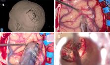
          We performed a complete resection of a thalamic/internal capsule CM using a tubular retractor system via a parietal trans-sulcal para-fascicular (PTPF) approach without collateral injury to the nearby white matter tracts.

          Conclusion

          PTPF approach to lateral thalamic/internal capsule lesions can be safely performed without injury to eloquent white matter fibres. The paucity of major vessels along this trajectory and the preservation of lateral ventricle integrity make this approach a feasible alternative to traditional approaches.

          Supplementary Information

          The online version contains supplementary material available at 10.1007/s00701-021-04884-2.

          Related collections

          Most cited references7

          • Record: found
          • Abstract: found
          • Article: not found

          Hemorrhage from cerebral cavernous malformations: a systematic pooled analysis.

          OBJECTIVE The aim of this paper is to define an overall cavernous malformation (CM) hemorrhage rate and risk factors for hemorrhage. METHODS The authors performed a systematic, pooled analysis via the PubMed database through October 2015 using the terms "cavernoma," "cavernous malformation," "natural history," "bleeding," and "hemorrhage." English-language studies providing annual rates and/or risk factors for CM hemorrhage were included. Data extraction, performed independently by the authors, included demographic data, hemorrhage rates, and hemorrhage risk factors. RESULTS Across 12 natural history studies with 1610 patients, the mean age at presentation was 42.7 years old and 52% of patients (95% CI 49%-55%) were female. Presentation modality was seizure in 30% (95% CI 25%-35%), hemorrhage in 26% (95% CI 17%-37%), incidental in 17% (95% CI 9%-31%), and focal deficits only in 16% of cases (95% CI 11%-23%). CM location was lobar in 66% (95% CI 61%-70%), brainstem in 18% (95% CI 13%-24%), deep supratentorial in 8% (95% CI 6%-10%), and cerebellar in 8% (95% CI 5%-11%). Pooling 7 studies that did not assume CM presence since birth, the annual hemorrhage rate was 2.5% per patient-year over 5081.2 patient-years of follow-up (95% CI 1.3%-5.1%). Pooling hazard ratios across 5 studies that evaluated hemorrhage risk factors, prior CM hemorrhage was a significant risk factor for hemorrhage (HR 3.73, 95% CI 1.26-11.1; p = 0.02) while younger age, female sex, deep location, size, multiplicity, and associated developmental venous anomalies (DVAs) were not. CONCLUSIONS Although limited by the heterogeneity of incorporated reports and selection bias, this study found prior hemorrhage to be a significant risk factor for CM bleeding, while age, sex, CM location, size, multiplicity, and associated DVAs were not. Future natural history studies should compound annual hemorrhage rate with prospective seizure and nonhemorrhagic neurological deficit rates.
            Bookmark
            • Record: found
            • Abstract: found
            • Article: not found

            Cerebral cavernous malformations: natural history and prognosis after clinical deterioration with or without hemorrhage.

            Despite recent studies of the natural history of cavernous malformations, there remains significant uncertainty concerning hemorrhage rates and the importance of lesion location. Controversy arises over varying definitions of "hemorrhage." What is ultimately important to the patient is the occurrence of a neurological event, which may or may not be associated with radiologically documented hemorrhage, as well as the chance of recovery after such an event. The purpose of this study was to determine the rates of occurrence and sequelae of neurological events in 173 patients referred to our vascular malformation clinic with cavernous malformations. All patient data were entered into a database. The mean age at presentation for the 173 patients was 37.5 years. The lesion location was deep (brainstem, cerebellar nuclei, thalamus, or basal ganglia) in 64 patients (37%) and superficial in 109 (63%). Thirty-one patients (18%) had multiple lesions. Disease presentation was due to seizures in 62 patients (36%), hemorrhage in 44 (25%), focal neurological deficit without documented hemorrhage in 35 (20%), headache alone in 11 (6%), and incidental findings in 21 patients (12%). The results obtained in the 110 patients eligible for follow-up review were used to derive information on the rates of hemorrhage and neurological events. An interval event (neurological deterioration) required both symptoms and signs. The total mean follow-up period was 46 months, the majority (65%) of which was prospective. There were 18 interval events in 427 patient-years of follow-up review, for an overall annual event rate of 4.2%. Location was the most important factor for predicting interval event occurrence, with significantly higher rates for deeply located (10.6%/year) compared with superficially located lesions (0%/year) (p = 0.0001). Of patients suffering a neurological event, only 37% had complete resolution of their deficits. This largely prospective study indicates that deep cavernous malformations carry a worse prognosis than superficial lesions with respect to annual rates of neurological deterioration. The alarming rate of adverse clinical events occurring in patients with deep lesions is punctuated by the fact that less than one-half of them recover fully during long-term follow-up review.
              Bookmark
              • Record: found
              • Abstract: not found
              • Article: not found

              Cranial Cavernous Malformations

                Bookmark

                Author and article information

                Contributors
                michaelamoo@rcsi.ie
                Journal
                Acta Neurochir (Wien)
                Acta Neurochir (Wien)
                Acta Neurochirurgica
                Springer Vienna (Vienna )
                0001-6268
                0942-0940
                24 June 2021
                24 June 2021
                2021
                : 163
                : 9
                : 2497-2501
                Affiliations
                [1 ]Department of Neurosurgery, Beacon Hospital, Sandyford, Dublin 18, Ireland
                [2 ]GRID grid.414315.6, ISNI 0000 0004 0617 6058, National Neurosurgical Centre, , Beaumont Hospital, ; Dublin 9, Ireland
                [3 ]GRID grid.4912.e, ISNI 0000 0004 0488 7120, Royal College of Surgeons in Ireland, ; Dublin, Ireland
                [4 ]GRID grid.414315.6, ISNI 0000 0004 0617 6058, Department of Neurology and Clinical Neurophysiology, , Beaumont Hospital, ; Dublin 9, Ireland
                [5 ]GRID grid.8217.c, ISNI 0000 0004 1936 9705, Department of Academic Neurology, , Trinity College Dublin, ; Dublin, Ireland
                Author information
                http://orcid.org/0000-0001-6886-6714
                http://orcid.org/0000-0001-9836-1741
                Article
                4884
                10.1007/s00701-021-04884-2
                8357681
                34164736
                d7e32759-3bde-4d1a-9462-4caa43492575
                © The Author(s) 2021

                Open AccessThis article is licensed under a Creative Commons Attribution 4.0 International License, which permits use, sharing, adaptation, distribution and reproduction in any medium or format, as long as you give appropriate credit to the original author(s) and the source, provide a link to the Creative Commons licence, and indicate if changes were made. The images or other third party material in this article are included in the article's Creative Commons licence, unless indicated otherwise in a credit line to the material. If material is not included in the article's Creative Commons licence and your intended use is not permitted by statutory regulation or exceeds the permitted use, you will need to obtain permission directly from the copyright holder. To view a copy of this licence, visit http://creativecommons.org/licenses/by/4.0/.

                History
                : 2 March 2021
                : 13 May 2021
                Funding
                Funded by: Royal College of Surgeons in Ireland (RCSI)
                Categories
                How I Do it - Vascular Neurosurgery - Other
                Custom metadata
                © Springer-Verlag GmbH Austria, part of Springer Nature 2021

                Surgery
                thalamus,internal capsule,brainpath®,tubular retractor,para-fascicular surgery,cavernous malformation,minimally invasive neurosurgery,exoscope,orbeye®

                Comments

                Comment on this article