12
views
0
recommends
+1 Recommend
0 collections
    0
    shares
      • Record: found
      • Abstract: found
      • Article: found
      Is Open Access

      Enhanced release of acid sphingomyelinase-enriched exosomes generates a lipidomics signature in CSF of Multiple Sclerosis patients

      research-article

      Read this article at

      Bookmark
          There is no author summary for this article yet. Authors can add summaries to their articles on ScienceOpen to make them more accessible to a non-specialist audience.

          Abstract

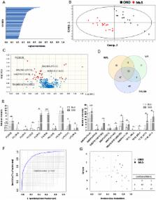
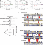
          Multiple Sclerosis (MuS) is a complex multifactorial neuropathology, resulting in heterogeneous clinical presentation. A very active MuS research field concerns the discovery of biomarkers helpful to make an early and definite diagnosis. The sphingomyelin pathway has emerged as a molecular mechanism involved in MuS, since high levels of ceramides in cerebrospinal fluid (CSF) were related to axonal damage and neuronal dysfunction. Ceramides are the hydrolysis products of sphingomyelins through a reaction catalyzed by a family of enzymes named sphingomyelinases, which were recently related to myelin repair in MuS. Here, using a lipidomic approach, we observed low levels of several sphingomyelins in CSF of MuS patients compared to other inflammatory and non-inflammatory, central or peripheral neurological diseases. Starting by this result, we investigated the sphingomyelinase activity in CSF, showing a significantly higher enzyme activity in MuS. In support of these results we found high number of total exosomes in CSF of MuS patients and a high number of acid sphingomyelinase-enriched exosomes correlated to enzymatic activity and to disease severity. These data are of diagnostic relevance and show, for the first time, high number of acid sphingomyelinase-enriched exosomes in MuS, opening a new window for therapeutic approaches/targets in the treatment of MuS.

          Related collections

          Most cited references25

          • Record: found
          • Abstract: found
          • Article: not found

          Deregulation of sphingolipid metabolism in Alzheimer's disease.

          Abnormal sphingolipid metabolism has been previously reported in Alzheimer's disease (AD). To extend these findings, several sphingolipids and sphingolipid hydrolases were analyzed in brain samples from AD patients and age-matched normal individuals. We found a pattern of elevated acid sphingomyelinase (ASM) and acid ceramidase (AC) expression in AD, leading to a reduction in sphingomyelin and elevation of ceramide. More sphingosine also was found in the AD brains, although sphingosine-1-phosphate (S1P) levels were reduced. Notably, significant correlations were observed between the brain ASM and S1P levels and the levels of amyloid beta (Abeta) peptide and hyperphosphorylated tau protein. Based on these findings, neuronal cell cultures were treated with Abeta oligomers, which were found to activate ASM, increase ceramide, and induce apoptosis. Pre-treatment of the neurons with purified, recombinant AC prevented the cells from undergoing Abeta-induced apoptosis. We propose that ASM activation is an important pathological event leading to AD, perhaps due to Abeta deposition. The downstream consequences of ASM activation are elevated ceramide, activation of ceramidases, and production of sphingosine. The reduced levels of S1P in the AD brain, together with elevated ceramide, likely contribute to the disease pathogenesis. Copyright 2008 Elsevier Inc. All rights reserved.
            Bookmark
            • Record: found
            • Abstract: found
            • Article: not found

            Global exosome transcriptome profiling reveals biomarkers for multiple sclerosis.

            Accumulating evidence supports a role for exosomes in immune regulation. In this study, we investigated the total circulating exosome transcriptome in relapsing-remitting multiple sclerosis (RRMS) patients and healthy controls (HC).
              Bookmark
              • Record: found
              • Abstract: found
              • Article: found
              Is Open Access

              A defect of sphingolipid metabolism modifies the properties of normal appearing white matter in multiple sclerosis

              Maintaining the appropriate complement and content of lipids in cellular membranes is critical for normal neural function. Accumulating evidence suggests that even subtle perturbations in the lipid content of neurons and myelin can disrupt their function and may contribute to myelin and axonal degradation. In this study, we determined the composition and quantified the content of lipids and sterols in normal appearing white matter (NAWM) and normal appearing grey matter (NAGM) from control and multiple sclerosis brain tissues by electrospray ionization tandem mass spectrometry. Our results suggest that in active-multiple sclerosis, there is a shift in the lipid composition of NAWM and NAGM to a higher phospholipid and lower sphingolipid content. We found that this disturbance in lipid composition was reduced in NAGM but not in NAWM of inactive-multiple sclerosis. The pattern of disturbance in lipid composition suggests a metabolic defect that causes sphingolipids to be shuttled to phospholipid production. Modelling the biophysical consequence of this change in lipid composition of NAWM indicated an increase in the repulsive force between opposing bilayers that could explain decompaction and disruption of myelin structure.
                Bookmark

                Author and article information

                Contributors
                piero.delboccio@unich.it
                Journal
                Sci Rep
                Sci Rep
                Scientific Reports
                Nature Publishing Group UK (London )
                2045-2322
                15 February 2018
                15 February 2018
                2018
                : 8
                : 3071
                Affiliations
                [1 ]ISNI 0000 0001 2181 4941, GRID grid.412451.7, Department of Medical, Oral and Biotechnological Sciences, , University “G. d’Annunzio” of Chieti-Pescara, ; Chieti, Italy
                [2 ]ISNI 0000 0001 2181 4941, GRID grid.412451.7, Centre on Aging Sciences and Translational Medicine (Ce.S.I-MeT), , University “G. d’Annunzio” of Chieti-Pescara, ; Chieti, Italy
                [3 ]ISNI 0000 0001 2181 4941, GRID grid.412451.7, Department of Pharmacy, , University “G. d’Annunzio” of Chieti-Pescara, ; Chieti, Italy
                [4 ]ISNI 0000 0001 2181 4941, GRID grid.412451.7, Department of Medicine and Aging Sciences, , “G. d’Annunzio” University of Chieti-Pescara, ; Chieti, Italy
                [5 ]ISNI 0000 0001 2181 4941, GRID grid.412451.7, Department of Neuroscience, Imaging and Clinical Sciences, , “G. d’Annunzio” University of Chieti-Pescara, ; Chieti, Italy
                Author information
                http://orcid.org/0000-0003-1015-3484
                http://orcid.org/0000-0003-1653-2194
                Article
                21497
                10.1038/s41598-018-21497-5
                5814401
                29449691
                d71b7153-a996-4086-b29a-6a7256437137
                © The Author(s) 2018

                Open Access This article is licensed under a Creative Commons Attribution 4.0 International License, which permits use, sharing, adaptation, distribution and reproduction in any medium or format, as long as you give appropriate credit to the original author(s) and the source, provide a link to the Creative Commons license, and indicate if changes were made. The images or other third party material in this article are included in the article’s Creative Commons license, unless indicated otherwise in a credit line to the material. If material is not included in the article’s Creative Commons license and your intended use is not permitted by statutory regulation or exceeds the permitted use, you will need to obtain permission directly from the copyright holder. To view a copy of this license, visit http://creativecommons.org/licenses/by/4.0/.

                History
                : 19 October 2017
                : 1 February 2018
                Categories
                Article
                Custom metadata
                © The Author(s) 2018

                Uncategorized
                Uncategorized

                Comments

                Comment on this article