3
views
0
recommends
+1 Recommend
0 collections
    0
    shares
      • Record: found
      • Abstract: found
      • Article: found
      Is Open Access

      Unveiling a novel memory center in human brain: neurochemical identification of the nucleus incertus, a key pontine locus implicated in stress and neuropathology

      research-article

      Read this article at

      Bookmark
          There is no author summary for this article yet. Authors can add summaries to their articles on ScienceOpen to make them more accessible to a non-specialist audience.

          Abstract

          Background

          The nucleus incertus (NI) was originally described by Streeter in 1903, as a midline region in the floor of the fourth ventricle of the human brain with an ‘unknown’ function. More than a century later, the neuroanatomy of the NI has been described in lower vertebrates, but not in humans. Therefore, we examined the neurochemical anatomy of the human NI using markers, including the neuropeptide, relaxin-3 (RLN3), and began to explore the distribution of the NI-related RLN3 innervation of the hippocampus.

          Methods

          Histochemical staining of serial, coronal sections of control human postmortem pons was conducted to reveal the presence of the NI by detection of immunoreactivity (IR) for the neuronal markers, microtubule-associated protein-2 (MAP2), glutamic acid dehydrogenase (GAD)-65/67 and corticotrophin-releasing hormone receptor 1 (CRHR1), and RLN3, which is highly expressed in NI neurons in diverse species. RLN3 and vesicular GABA transporter 1 ( vGAT1) mRNA were detected by fluorescent in situ hybridization. Pons sections containing the NI from an AD case were immunostained for phosphorylated-tau, to explore potential relevance to neurodegenerative diseases. Lastly, sections of the human hippocampus were stained to detect RLN3-IR and somatostatin (SST)-IR.

          Results

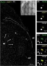
          In the dorsal, anterior-medial region of the human pons, neurons containing RLN3- and MAP2-IR, and RLN3/vGAT1 mRNA-positive neurons were observed in an anatomical pattern consistent with that of the NI in other species. GAD65/67- and CRHR1-immunopositive neurons were also detected within this area. Furthermore, RLN3- and AT8-IR were co-localized within NI neurons of an AD subject. Lastly, RLN3-IR was detected in neurons within the CA1, CA2, CA3 and DG areas of the hippocampus, in the absence of RLN3 mRNA. In the DG, RLN3- and SST-IR were co-localized in a small population of neurons.

          Conclusions

          Aspects of the anatomy of the human NI are shared across species, including a population of stress-responsive, RLN3-expressing neurons and a RLN3 innervation of the hippocampus. Accumulation of phosphorylated-tau in the NI suggests its possible involvement in AD pathology. Further characterization of the neurochemistry of the human NI will increase our understanding of its functional role in health and disease.

          Graphical Abstract

          Supplementary Information

          The online version contains supplementary material available at 10.1186/s40659-024-00523-z.

          Related collections

          Most cited references93

          • Record: found
          • Abstract: found
          • Article: not found

          NIH Image to ImageJ: 25 years of image analysis

          For the past twenty five years the NIH family of imaging software, NIH Image and ImageJ have been pioneers as open tools for scientific image analysis. We discuss the origins, challenges and solutions of these two programs, and how their history can serve to advise and inform other software projects.
            Bookmark
            • Record: found
            • Abstract: found
            • Article: not found

            Stages of the pathologic process in Alzheimer disease: age categories from 1 to 100 years.

            Two thousand three hundred and thirty two nonselected brains from 1- to 100-year-old individuals were examined using immunocytochemistry (AT8) and Gallyas silver staining for abnormal tau; immunocytochemistry (4G8) and Campbell-Switzer staining were used for the detection ofβ-amyloid. A total of 342 cases was negative in the Gallyas stain but when restaged for AT8 only 10 were immunonegative. Fifty-eight cases had subcortical tau predominantly in the locus coeruleus, but there was no abnormal cortical tau (subcortical Stages a-c). Cortical involvement (abnormal tau in neurites) was identified first in the transentorhinal region (Stage 1a, 38 cases). Transentorhinal pyramidal cells displayed pretangle material (Stage 1b, 236 cases). Pretangles gradually became argyrophilic neurofibrillary tangles (NFTs) that progressed in parallel with NFT Stages I to VI. Pretangles restricted to subcortical sites were seen chiefly at younger ages. Of the total cases, 1,031 (44.2%) had β-amyloid plaques. The first plaques occurred in the neocortex after the onset of tauopathy in the brainstem. Plaques generally developed in the 40s in 4% of all cases, culminating in their tenth decade (75%). β-amyloid plaques and NFTs were significantly correlated (p < 0.0001). These data suggest that tauopathy associated with sporadic Alzheimer disease may begin earlier than previously thought and possibly in the lower brainstem rather than in the transentorhinal region.
              Bookmark
              • Record: found
              • Abstract: found
              • Article: not found

              Tau protein isoforms, phosphorylation and role in neurodegenerative disorders.

              Tau proteins belong to the family of microtubule-associated proteins. They are mainly expressed in neurons where they play an important role in the assembly of tubulin monomers into microtubules to constitute the neuronal microtubules network. Microtubules are involved in maintaining the cell shape and serve as tracks for axonal transport. Tau proteins also establish some links between microtubules and other cytoskeletal elements or proteins. Tau proteins are translated from a single gene located on chromosome 17. Their expression is developmentally regulated by an alternative splicing mechanism and six different isoforms exist in the human adult brain. Tau proteins are the major constituents of intraneuronal and glial fibrillar lesions described in Alzheimer's disease and numerous neurodegenerative disorders referred to as 'tauopathies'. Molecular analysis has revealed that an abnormal phosphorylation might be one of the important events in the process leading to their aggregation. Moreover, a specific set of pathological tau proteins exhibiting a typical biochemical pattern, and a different regional and laminar distribution could characterize each of these disorders. Finally, a direct correlation has been established between the progressive involvement of the neocortical areas and the increasing severity of dementia, suggesting that pathological tau proteins are reliable marker of the neurodegenerative process. The recent discovery of tau gene mutations in frontotemporal dementia with parkinsonism linked to chromosome 17 has reinforced the predominant role attributed to tau proteins in the pathogenesis of neurodegenerative disorders, and underlined the fact that distinct sets of tau isoforms expressed in different neuronal populations could lead to different pathologies.
                Bookmark

                Author and article information

                Contributors
                cdeavila@asu.edu
                Journal
                Biol Res
                Biol Res
                Biological Research
                BioMed Central (London )
                0716-9760
                0717-6287
                16 July 2024
                16 July 2024
                2024
                : 57
                : 46
                Affiliations
                [1 ]Arizona State University-Banner Neurodegenerative Disease Research Center, ( https://ror.org/03efmqc40) Tempe, AZ USA
                [2 ]Department of Neurophysiology and Chronobiology, Institute of Zoology and Biomedical Research, Jagiellonian University, ( https://ror.org/03bqmcz70) Krakow, Poland
                [3 ]Arizona Alzheimer’s Consortium, ( https://ror.org/00cvnc278) Phoenix, AZ USA
                [4 ]Banner Sun Health Research Institute, ( https://ror.org/04gjkkf30) Sun City, AZ USA
                [5 ]GRID grid.421142.0, ISNI 0000 0000 8521 1798, Quebec Heart and Lung Institute, ; Quebec City, QC Canada
                [6 ]Department of Neuroscience, Mayo Clinic, ( https://ror.org/02qp3tb03) Scottsdale, AZ USA
                [7 ]Faculty of Pharmacy, Université Laval, ( https://ror.org/04sjchr03) Quebec City, QC Canada
                [8 ]GRID grid.1008.9, ISNI 0000 0001 2179 088X, Florey Department of Neuroscience and Mental Health and Department of Anatomy and Physiology and The Florey Institute of Neuroscience and Mental Health, , The University of Melbourne, ; Melbourne, VIC Australia
                Author information
                http://orcid.org/0000-0003-1067-5952
                Article
                523
                10.1186/s40659-024-00523-z
                11253401
                39014514
                d7162262-d1fe-4fcf-b236-8af293c26c98
                © The Author(s) 2024

                Open Access This article is licensed under a Creative Commons Attribution 4.0 International License, which permits use, sharing, adaptation, distribution and reproduction in any medium or format, as long as you give appropriate credit to the original author(s) and the source, provide a link to the Creative Commons licence, and indicate if changes were made. The images or other third party material in this article are included in the article's Creative Commons licence, unless indicated otherwise in a credit line to the material. If material is not included in the article's Creative Commons licence and your intended use is not permitted by statutory regulation or exceeds the permitted use, you will need to obtain permission directly from the copyright holder. To view a copy of this licence, visit http://creativecommons.org/licenses/by/4.0/. The Creative Commons Public Domain Dedication waiver ( http://creativecommons.org/publicdomain/zero/1.0/) applies to the data made available in this article, unless otherwise stated in a credit line to the data.

                History
                : 13 October 2023
                : 7 June 2024
                Funding
                Funded by: FundRef http://dx.doi.org/10.13039/100000065, National Institute of Neurological Disorders and Stroke;
                Award ID: U24 NS072026 National Brain
                Award ID: Tissue Resource for Parkinson’s Disease
                Award ID: Related Disorders
                Funded by: FundRef http://dx.doi.org/10.13039/100000049, National Institute on Aging;
                Award ID: P30 AG19610 Arizona Alzheimer’s Disease Core Center
                Funded by: Arizona Department of Health Services
                Award ID: contract 211002
                Award ID: Arizona Alzheimer’s Research Center
                Funded by: FundRef http://dx.doi.org/10.13039/100008335, Arizona Biomedical Research Commission;
                Award ID: contracts 4001
                Award ID: 0011
                Award ID: 05-901
                Award ID: 1001 to the Arizona Parkinson's Disease Consortium
                Funded by: FundRef http://dx.doi.org/10.13039/100000864, Michael J. Fox Foundation for Parkinson's Research;
                Funded by: Strategic Program Initiative of Excellence in the Jagiellonian University
                Award ID: Minigrant at the Faculty of Biology 2023
                Award Recipient :
                Funded by: FundRef http://dx.doi.org/10.13039/100006312, BrightFocus Foundation;
                Award ID: A2021006
                Award Recipient :
                Funded by: FundRef http://dx.doi.org/10.13039/100000957, Alzheimer's Association;
                Award ID: AARFD-22-972099
                Award Recipient :
                Categories
                Research Article
                Custom metadata
                © Sociedad de Biologia de Chile 2024

                brainstem,dementia,hippocampus,human,memory,nucleus incertus,relaxin-3

                Comments

                Comment on this article