123
views
0
recommends
+1 Recommend
0 collections
    0
    shares
      • Record: found
      • Abstract: found
      • Article: found
      Is Open Access

      Microglia protect against brain injury and their selective elimination dysregulates neuronal network activity after stroke

      research-article

      Read this article at

      Bookmark
          There is no author summary for this article yet. Authors can add summaries to their articles on ScienceOpen to make them more accessible to a non-specialist audience.

          Abstract

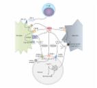
          Microglia are the main immune cells of the brain and contribute to common brain diseases. However, it is unclear how microglia influence neuronal activity and survival in the injured brain in vivo. Here we develop a precisely controlled model of brain injury induced by cerebral ischaemia combined with fast in vivo two-photon calcium imaging and selective microglial manipulation. We show that selective elimination of microglia leads to a striking, 60% increase in infarct size, which is reversed by microglial repopulation. Microglia-mediated protection includes reduction of excitotoxic injury, since an absence of microglia leads to dysregulated neuronal calcium responses, calcium overload and increased neuronal death. Furthermore, the incidence of spreading depolarization (SD) is markedly reduced in the absence of microglia. Thus, microglia are involved in changes in neuronal network activity and SD after brain injury in vivo that could have important implications for common brain diseases.

          Abstract

          How microglia contribute to brain injury or repair is unclear. Here combining microglia manipulations and calcium imaging, the authors show that selective elimination of microglia leads to disrupted neuronal calcium dynamics and markedly increased brain injury after cerebral ischemia.

          Related collections

          Most cited references36

          • Record: found
          • Abstract: found
          • Article: not found

          The role of spreading depression, spreading depolarization and spreading ischemia in neurological disease.

          The term spreading depolarization describes a wave in the gray matter of the central nervous system characterized by swelling of neurons, distortion of dendritic spines, a large change of the slow electrical potential and silencing of brain electrical activity (spreading depression). In the clinic, unequivocal electrophysiological evidence now exists that spreading depolarizations occur abundantly in individuals with aneurismal subarachnoid hemorrhage, delayed ischemic stroke after subarachnoid hemorrhage, malignant hemispheric stroke, spontaneous intracerebral hemorrhage or traumatic brain injury. Spreading depolarization is induced experimentally by various noxious conditions including chemicals such as potassium, glutamate, inhibitors of the sodium pump, status epilepticus, hypoxia, hypoglycemia and ischemia, but it can can also invade healthy, naive tissue. Resistance vessels respond to it with tone alterations, causing either transient hyperperfusion (physiological hemodynamic response) in healthy tissue or severe hypoperfusion (inverse hemodynamic response, or spreading ischemia) in tissue at risk for progressive damage, which contributes to lesion progression. Therapies that target spreading depolarization or the inverse hemodynamic response may potentially treat these neurological conditions.
            Bookmark
            • Record: found
            • Abstract: found
            • Article: not found

            Superresolution imaging of chemical synapses in the brain.

            Determination of the molecular architecture of synapses requires nanoscopic image resolution and specific molecular recognition, a task that has so far defied many conventional imaging approaches. Here, we present a superresolution fluorescence imaging method to visualize the molecular architecture of synapses in the brain. Using multicolor, three-dimensional stochastic optical reconstruction microscopy, the distributions of synaptic proteins can be measured with nanometer precision. Furthermore, the wide-field, volumetric imaging method enables high-throughput, quantitative analysis of a large number of synapses from different brain regions. To demonstrate the capabilities of this approach, we have determined the organization of ten protein components of the presynaptic active zone and the postsynaptic density. Variations in synapse morphology, neurotransmitter receptor composition, and receptor distribution were observed both among synapses and across different brain regions. Combination with optogenetics further allowed molecular events associated with synaptic plasticity to be resolved at the single-synapse level. Copyright © 2010 Elsevier Inc. All rights reserved.
              Bookmark
              • Record: found
              • Abstract: found
              • Article: found
              Is Open Access

              Tumor Necrosis Factor Alpha: A Link between Neuroinflammation and Excitotoxicity

              Tumor necrosis factor alpha (TNF- α ) is a proinflammatory cytokine that exerts both homeostatic and pathophysiological roles in the central nervous system. In pathological conditions, microglia release large amounts of TNF- α ; this de novo production of TNF- α is an important component of the so-called neuroinflammatory response that is associated with several neurological disorders. In addition, TNF- α can potentiate glutamate-mediated cytotoxicity by two complementary mechanisms: indirectly, by inhibiting glutamate transport on astrocytes, and directly, by rapidly triggering the surface expression of Ca+2 permeable-AMPA receptors and NMDA receptors, while decreasing inhibitory GABAA receptors on neurons. Thus, the net effect of TNF- α is to alter the balance of excitation and inhibition resulting in a higher synaptic excitatory/inhibitory ratio. This review summarizes the current knowledge of the cellular and molecular mechanisms by which TNF- α links the neuroinflammatory and excitotoxic processes that occur in several neurodegenerative diseases, but with a special emphasis on amyotrophic lateral sclerosis (ALS). As microglial activation and upregulation of TNF- α expression is a common feature of several CNS diseases, as well as chronic opioid exposure and neuropathic pain, modulating TNF- α signaling may represent a valuable target for intervention.
                Bookmark

                Author and article information

                Journal
                Nat Commun
                Nat Commun
                Nature Communications
                Nature Publishing Group
                2041-1723
                03 May 2016
                2016
                : 7
                : 11499
                Affiliations
                [1 ]Two-Photon Imaging Center, Institute of Experimental Medicine, Hungarian Academy of Sciences , Szigony U. 43, Budapest 1083, Hungary
                [2 ]Laboratory of Neuroimmunology, Institute of Experimental Medicine, Hungarian Academy of Sciences , Szigony U. 43, Budapest 1083, Hungary
                [3 ]MTA-PPKE ITK-NAP B - Two-photon measurement Technology Research Group, Pázmány Péter University , Budapest 1083, Hungary
                [4 ]Plexxikon, Inc. , Berkeley, California 94710, USA
                Author notes
                [*]

                These authors contributed equally to this work.

                [†]

                These authors jointly supervised this work.

                Article
                ncomms11499
                10.1038/ncomms11499
                4857403
                27139776
                d5b150d3-04f1-4cd9-94c8-a49a8fc4073c
                Copyright © 2016, Nature Publishing Group, a division of Macmillan Publishers Limited. All Rights Reserved.

                This work is licensed under a Creative Commons Attribution 4.0 International License. The images or other third party material in this article are included in the article's Creative Commons license, unless indicated otherwise in the credit line; if the material is not included under the Creative Commons license, users will need to obtain permission from the license holder to reproduce the material. To view a copy of this license, visit http://creativecommons.org/licenses/by/4.0/

                History
                : 26 November 2015
                : 04 April 2016
                Categories
                Article

                Uncategorized
                Uncategorized

                Comments

                Comment on this article