11
views
0
recommends
+1 Recommend
0 collections
    0
    shares
      • Record: found
      • Abstract: found
      • Article: not found

      A large-scale standardized physiological survey reveals functional organization of the mouse visual cortex

      research-article
      1 , # , 1 , # , 1 , # , 1 , 1 , 1 , 1 , 1 , 1 , 1 , 1 , 1 , 1 , 1 , 1 , 1 , 1 , 1 , 1 , 1 , 1 , 1 , 1 , 1 , 1 , 1 , 1 , 1 , 1 , 1 , 1 , 1 , 1 , 1 , 1 , 1 , 1 , 1 , 2 , 1 , 1 , 1 , 1 , 1 , 1 , 1 , 1 , 1 , 1 , 1 , 1 , 1 , 1 , 1 , 1 , 1 , 2 , 2 , 1 , 1 , 1 , 1 , 1 , 1 , 2 , 1 , 1 , 1 , 1 , 1 , 1 , 1 , 1
      Nature neuroscience

      Read this article at

      ScienceOpenPublisherPMC
      Bookmark
          There is no author summary for this article yet. Authors can add summaries to their articles on ScienceOpen to make them more accessible to a non-specialist audience.

          Summary

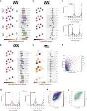
          To understand how the brain processes sensory information to guide behavior, we must know how stimulus representations are transformed throughout the visual cortex. Here we report an open, large-scale physiological survey of activity in the awake mouse visual cortex: the Allen Brain Observatory Visual Coding dataset. This publicly available dataset includes cortical activity from nearly 60,000 neurons from 6 visual areas, 4 layers, and 12 transgenic mouse lines from 243 adult mice, in response to a systematic set of visual stimuli. We classify neurons based on joint reliabilities to multiple stimuli and validate this functional classification with models of visual responses. While most classes are characterized by responses to specific subsets of the stimuli, the largest class is not reliably responsive to any of the stimuli and becomes progressively larger in higher visual areas. These classes reveal a functional organization wherein putative dorsal areas show specialization for visual motion signals.

          Related collections

          Most cited references68

          • Record: found
          • Abstract: found
          • Article: not found

          A mesoscale connectome of the mouse brain.

          Comprehensive knowledge of the brain's wiring diagram is fundamental for understanding how the nervous system processes information at both local and global scales. However, with the singular exception of the C. elegans microscale connectome, there are no complete connectivity data sets in other species. Here we report a brain-wide, cellular-level, mesoscale connectome for the mouse. The Allen Mouse Brain Connectivity Atlas uses enhanced green fluorescent protein (EGFP)-expressing adeno-associated viral vectors to trace axonal projections from defined regions and cell types, and high-throughput serial two-photon tomography to image the EGFP-labelled axons throughout the brain. This systematic and standardized approach allows spatial registration of individual experiments into a common three dimensional (3D) reference space, resulting in a whole-brain connectivity matrix. A computational model yields insights into connectional strength distribution, symmetry and other network properties. Virtual tractography illustrates 3D topography among interconnected regions. Cortico-thalamic pathway analysis demonstrates segregation and integration of parallel pathways. The Allen Mouse Brain Connectivity Atlas is a freely available, foundational resource for structural and functional investigations into the neural circuits that support behavioural and cognitive processes in health and disease.
            Bookmark
            • Record: found
            • Abstract: found
            • Article: not found

            A resource of Cre driver lines for genetic targeting of GABAergic neurons in cerebral cortex.

            A key obstacle to understanding neural circuits in the cerebral cortex is that of unraveling the diversity of GABAergic interneurons. This diversity poses general questions for neural circuit analysis: how are these interneuron cell types generated and assembled into stereotyped local circuits and how do they differentially contribute to circuit operations that underlie cortical functions ranging from perception to cognition? Using genetic engineering in mice, we have generated and characterized approximately 20 Cre and inducible CreER knockin driver lines that reliably target major classes and lineages of GABAergic neurons. More select populations are captured by intersection of Cre and Flp drivers. Genetic targeting allows reliable identification, monitoring, and manipulation of cortical GABAergic neurons, thereby enabling a systematic and comprehensive analysis from cell fate specification, migration, and connectivity, to their functions in network dynamics and behavior. As such, this approach will accelerate the study of GABAergic circuits throughout the mammalian brain. Copyright © 2011 Elsevier Inc. All rights reserved.
              Bookmark
              • Record: found
              • Abstract: found
              • Article: not found

              Fully integrated silicon probes for high-density recording of neural activity

              Sensory, motor and cognitive operations involve the coordinated action of large neuronal populations across multiple brain regions in both superficial and deep structures. Existing extracellular probes record neural activity with excellent spatial and temporal (sub-millisecond) resolution, but from only a few dozen neurons per shank. Optical Ca2+ imaging offers more coverage but lacks the temporal resolution needed to distinguish individual spikes reliably and does not measure local field potentials. Until now, no technology compatible with use in unrestrained animals has combined high spatiotemporal resolution with large volume coverage. Here we design, fabricate and test a new silicon probe known as Neuropixels to meet this need. Each probe has 384 recording channels that can programmably address 960 complementary metal–oxide–semiconductor (CMOS) processing-compatible low-impedance TiN sites that tile a single 10-mm long, 70 × 20-μm cross-section shank. The 6 × 9-mm probe base is fabricated with the shank on a single chip. Voltage signals are filtered, amplified, multiplexed and digitized on the base, allowing the direct transmission of noise-free digital data from the probe. The combination of dense recording sites and high channel count yielded well-isolated spiking activity from hundreds of neurons per probe implanted in mice and rats. Using two probes, more than 700 well-isolated single neurons were recorded simultaneously from five brain structures in an awake mouse. The fully integrated functionality and small size of Neuropixels probes allowed large populations of neurons from several brain structures to be recorded in freely moving animals. This combination of high-performance electrode technology and scalable chip fabrication methods opens a path towards recording of brain-wide neural activity during behaviour.
                Bookmark

                Author and article information

                Journal
                9809671
                21092
                Nat Neurosci
                Nat. Neurosci.
                Nature neuroscience
                1097-6256
                1546-1726
                13 November 2019
                16 December 2019
                January 2020
                16 June 2020
                : 23
                : 1
                : 138-151
                Affiliations
                [1 ]Allen Institute for Brain Science, Seattle, WA, USA
                [2 ]University of Washington, Seattle, WA, USA
                Author notes
                [*]

                These authors contributed equally to this work

                Author Contributions

                SEJdV, MAB, KR, MG, TK, SM, SO, JW, HZ, CD, LN, AB, JWP, RCR, and CK conceived of and designed the experiment. JAL, TK, PH, AL, CS, DS, and CF built and maintained the hardware. SEJdV, JAL, MAB, GKO, DF, NC, LK, WW, DW, RV, CB, BB, TD, JG, TG, SJ, NK, CL, F. Lee, F. Long, JP, NS, DMW, JZ, and LN developed algorithms and software, including the SDK and website. KR, N. Berbesque, N. Bowles, SC, LC, AC, SDC, ME, NG, FG, RH, LH, UK, JL, JDL, RL, EL, LL, JL, KM, TN, MR, SS, CW, and AW collected data. JAL, PAG, SEJdV, MAB supervised the work. SEJdV, JAL, MAB, GKO, MO, NC, PL, DM, JS, ESB, and RV analyzed data. CT and WW provided project administration. SEJdV, JAL, and MAB wrote the paper with input from PAG, GKO, MO, NC, PL, DM, RCR, MG, and CK.

                [# ]correspondence should be addressed to S.E.J.dV. ( saskiad@ 123456alleninstitute.org ), J.L. ( jeromel@ 123456alleninstitute.org ), and M.A.B. ( michaelbu@ 123456alleninstitute.org ).
                Article
                NIHMS1541598
                10.1038/s41593-019-0550-9
                6948932
                31844315
                d595bc95-aa36-4d1d-b1c4-2b6510484b6e

                Users may view, print, copy, and download text and data-mine the content in such documents, for the purposes of academic research, subject always to the full Conditions of use: http://www.nature.com/authors/editorial_policies/license.html#terms

                History
                Categories
                Article

                Neurosciences
                Neurosciences

                Comments

                Comment on this article