109
views
0
recommends
+1 Recommend
0 collections
    0
    shares
      • Record: found
      • Abstract: found
      • Article: found
      Is Open Access

      LPS-preconditioned mesenchymal stromal cells modify macrophage polarization for resolution of chronic inflammation via exosome-shuttled let-7b

      research-article

      Read this article at

      Bookmark
          There is no author summary for this article yet. Authors can add summaries to their articles on ScienceOpen to make them more accessible to a non-specialist audience.

          Abstract

          Background

          Within the last few years, it has become evident that LPS-preconditioned mesenchymal stromal cells (LPS pre-MSCs) show enhanced paracrine effects, including increased trophic support and improved regenerative and repair properties. MSCs may release large amounts of exosomes for cell-to-cell communication and maintain a dynamic and homeostatic microenvironment for tissue repair. The present study assesses the therapeutic efficacy and mechanisms of LPS-preconditioned MSC-derived exosomes (LPS pre-Exo) for chronic inflammation and wound healing.

          Methods

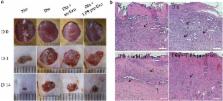
          We extracted exosomes from the supernatant of LPS pre-MSCs using a gradient centrifugation method. In vitro, THP-1 cells were cultured with high glucose (HG, 30 mM) as an inflammatory model and treated with LPS pre-Exo for 48 h. The expression of inflammation-related cytokines was detected by real-time RT-PCR, and the distribution of macrophage subtype was measured by immunofluorescence. Next, the miRNA expression profiles of LPS pre-Exo were evaluated using miRNA microarray analysis. The molecular signaling pathway responsible for the regenerative potential was identified by western blotting. In vivo, we established a cutaneous wound model in streptozotocin-induced diabetic rats, and LPS pre-Exo were injected dispersively into the wound edge. The curative effects of LPS pre-Exo on inflammation and wound healing were observed and evaluated.

          Results

          LPS pre-Exo have a better ability than untreated MSC-derived exosomes (un-Exo) to modulate the balance of macrophages due to their upregulation of the expression of anti-inflammatory cytokines and promotion of M2 macrophage activation. Microarray analysis of LPS pre-Exo identified the unique expression of let-7b compared with un-Exo, and the let-7b/TLR4 pathway served as potential contributor to macrophage polarization and inflammatory ablation. Further investigation of the mechanisms that control let-7b expression demonstrated that a TLR4/NF-κB/STAT3/AKT regulatory signaling pathway plays a critical role in the regulation of macrophage plasticity. Knockdown of AKT in THP-1 cells similarly abolished the immunomodulatory effect of LPS pre-Exo. In vivo, LPS pre-Exo greatly alleviated inflammation and enhanced diabetic cutaneous wound healing.

          Conclusion

          LPS pre-Exo may have improved regulatory abilities for macrophage polarization and resolution of chronic inflammation by shuttling let-7b, and these exosomes carry much immunotherapeutic potential for wound healing.

          Electronic supplementary material

          The online version of this article (doi:10.1186/s12967-015-0642-6) contains supplementary material, which is available to authorized users.

          Related collections

          Most cited references37

          • Record: found
          • Abstract: found
          • Article: found
          Is Open Access

          Exosomes released from human induced pluripotent stem cells-derived MSCs facilitate cutaneous wound healing by promoting collagen synthesis and angiogenesis

          Background Human induced pluripotent stem cell-derived mesenchymal stem cells (hiPSC-MSCs) have emerged as a promising alternative for stem cell transplantation therapy. Exosomes derived from mesenchymal stem cells (MSC-Exos) can play important roles in repairing injured tissues. However, to date, no reports have demonstrated the use of hiPSC-MSC-Exos in cutaneous wound healing, and little is known regarding their underlying mechanisms in tissue repair. Methods hiPSC-MSC-Exos were injected subcutaneously around wound sites in a rat model and the efficacy of hiPSC-MSC-Exos was assessed by measuring wound closure areas, by histological and immunofluorescence examinations. We also evaluated the in vitro effects of hiPSC-MSC-Exos on both the proliferation and migration of human dermal fibroblasts and human umbilical vein endothelial cells (HUVECs) by cell-counting and scratch assays, respectively. The effects of exosomes on fibroblast collagen and elastin secretion were studied in enzyme-linked immunosorbent assays and quantitative reverse-transcriptase–polymerase chain reaction (qRT-PCR). In vitro capillary network formation was determined in tube-formation assays. Results Transplanting hiPSC-MSC-Exos to wound sites resulted in accelerated re-epithelialization, reduced scar widths, and the promotion of collagen maturity. Moreover, hiPSC-MSC-Exos not only promoted the generation of newly formed vessels, but also accelerated their maturation in wound sites. We found that hiPSC-MSC-Exos stimulated the proliferation and migration of human dermal fibroblasts and HUVECs in a dose-dependent manner in vitro. Similarly, Type I, III collagen and elastin secretion and mRNA expression by fibroblasts and tube formation by HUVECs were also increased with increasing hiPSC-MSC-Exos concentrations. Conclusions Our findings suggest that hiPSC-MSC-Exos can facilitate cutaneous wound healing by promoting collagen synthesis and angiogenesis. These data provide the first evidence for the potential of hiPSC-MSC-Exos in treating cutaneous wounds.
            Bookmark
            • Record: found
            • Abstract: found
            • Article: found
            Is Open Access

            Mesenchymal stem cells reciprocally regulate the M1/M2 balance in mouse bone marrow-derived macrophages

            Mesenchymal stem cells (MSCs) have been widely studied for their applications in stem cell-based regeneration. During myocardial infarction (MI), infiltrated macrophages have pivotal roles in inflammation, angiogenesis and cardiac remodeling. We hypothesized that MSCs may modulate the immunologic environment to accelerate regeneration. This study was designed to assess the functional relationship between the macrophage phenotype and MSCs. MSCs isolated from bone marrow and bone marrow-derived macrophages (BMDMs) underwent differentiation induced by macrophage colony-stimulating factor. To determine the macrophage phenotype, classical M1 markers and alternative M2 markers were analyzed with or without co-culturing with MSCs in a transwell system. For animal studies, MI was induced by the ligation of the rat coronary artery. MSCs were injected within the infarct myocardium, and we analyzed the phenotype of the infiltrated macrophages by immunostaining. In the MSC-injected myocardium, the macrophages adjacent to the MSCs showed strong expression of arginase-1 (Arg1), an M2 marker. In BMDMs co-cultured with MSCs, the M1 markers such as interleukin-6 (IL-6), IL-1β, monocyte chemoattractant protein-1 and inducible nitric oxide synthase (iNOS) were significantly reduced. In contrast, the M2 markers such as IL-10, IL-4, CD206 and Arg1 were markedly increased by co-culturing with MSCs. Specifically, the ratio of iNOS to Arg1 in BMDMs was notably downregulated by co-culturing with MSCs. These results suggest that the preferential shift of the macrophage phenotype from M1 to M2 may be related to the immune-modulating characteristics of MSCs that contribute to cardiac repair.
              Bookmark
              • Record: found
              • Abstract: found
              • Article: not found

              The kinase Akt1 controls macrophage response to lipopolysaccharide by regulating microRNAs.

              MicroRNAs regulated by lipopolysaccharide (LPS) target genes that contribute to the inflammatory phenotype. Here, we showed that the protein kinase Akt1, which is activated by LPS, positively regulated miRNAs let-7e and miR-181c but negatively regulated miR-155 and miR-125b. In silico analyses and transfection studies revealed that let-7e repressed Toll-like receptor 4 (TLR4), whereas miR-155 repressed SOCS1, two proteins critical for LPS-driven TLR signaling, which regulate endotoxin sensitivity and tolerance. As a result, Akt1(-/-) macrophages exhibited increased responsiveness to LPS in culture and Akt1(-/-) mice did not develop endotoxin tolerance in vivo. Overexpression of let-7e and suppression of miR-155 in Akt1(-/-) macrophages restored sensitivity and tolerance to LPS in culture and in animals. These results indicate that Akt1 regulates the response of macrophages to LPS by controlling miRNA expression.
                Bookmark

                Author and article information

                Contributors
                tddee@163.com
                haojieh@163.com
                tc6636@sina.com
                ljj0558@126.com
                dl505504@qq.com
                zhengjxi@163.com
                zhaoyl301@163.com
                liu_hui_ling@sohu.com
                86-10-66937463 , fuxiaobing@vip.sina.com
                86-10-66937463 , hanwdrsw69@yahoo.com
                Journal
                J Transl Med
                J Transl Med
                Journal of Translational Medicine
                BioMed Central (London )
                1479-5876
                19 September 2015
                19 September 2015
                2015
                : 13
                : 308
                Affiliations
                [ ]Institute of Basic Medicine Science, College of Life Science, Chinese PLA General Hospital, No. 28 Fuxing Road, Beijing, 100853 China
                [ ]Central Laboratory, Hainan Branch of Chinese PLA General Hospital, Sanya, 572013 China
                Article
                642
                10.1186/s12967-015-0642-6
                4575470
                26386558
                d58768ce-451e-4087-9776-084c18e5ac32
                © Ti et al. 2015

                Open AccessThis article is distributed under the terms of the Creative Commons Attribution 4.0 International License ( http://creativecommons.org/licenses/by/4.0/), which permits unrestricted use, distribution, and reproduction in any medium, provided you give appropriate credit to the original author(s) and the source, provide a link to the Creative Commons license, and indicate if changes were made. The Creative Commons Public Domain Dedication waiver ( http://creativecommons.org/publicdomain/zero/1.0/) applies to the data made available in this article, unless otherwise stated.

                History
                : 3 July 2015
                : 18 August 2015
                Categories
                Research
                Custom metadata
                © The Author(s) 2015

                Medicine
                mesenchymal stromal cells,macrophage polarization,exosome,lps preconditioning
                Medicine
                mesenchymal stromal cells, macrophage polarization, exosome, lps preconditioning

                Comments

                Comment on this article