17
views
0
recommends
+1 Recommend
0 collections
    0
    shares
      • Record: found
      • Abstract: found
      • Article: found
      Is Open Access

      Retinal Vasculopathy in Alzheimer’s Disease

      review-article

      Read this article at

      Bookmark
          There is no author summary for this article yet. Authors can add summaries to their articles on ScienceOpen to make them more accessible to a non-specialist audience.

          Abstract

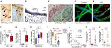
          The retina has been increasingly investigated as a site of Alzheimer’s disease (AD) manifestation for over a decade. Early reports documented degeneration of retinal ganglion cells and their axonal projections. Our group provided the first evidence of the key pathological hallmarks of AD, amyloid β-protein (Aβ) plaques including vascular Aβ deposits, in the retina of AD and mild cognitively impaired (MCI) patients. Subsequent studies validated these findings and further identified electroretinography and vision deficits, retinal (p)tau and inflammation, intracellular Aβ accumulation, and retinal ganglion cell-subtype degeneration surrounding Aβ plaques in these patients. Our data suggest that the brain and retina follow a similar trajectory during AD progression, probably due to their common embryonic origin and anatomical proximity. However, the retina is the only CNS organ feasible for direct, repeated, and non-invasive ophthalmic examination with ultra-high spatial resolution and sensitivity. Neurovascular unit integrity is key to maintaining normal CNS function and cerebral vascular abnormalities are increasingly recognized as early and pivotal factors driving cognitive impairment in AD. Likewise, retinal vascular abnormalities such as changes in vessel density and fractal dimensions, blood flow, foveal avascular zone, curvature tortuosity, and arteriole-to-venule ratio were described in AD patients including early-stage cases. A rapidly growing number of reports have suggested that cerebral and retinal vasculopathy are tightly associated with cognitive deficits in AD patients and animal models. Importantly, we recently identified early and progressive deficiency in retinal vascular platelet-derived growth factor receptor-β (PDGFRβ) expression and pericyte loss that were associated with retinal vascular amyloidosis and cerebral amyloid angiopathy in MCI and AD patients. Other studies utilizing optical coherence tomography (OCT), retinal amyloid-fluorescence imaging and retinal hyperspectral imaging have made significant progress in visualizing and quantifying AD pathology through the retina. With new advances in OCT angiography, OCT leakage, scanning laser microscopy, fluorescein angiography and adaptive optics imaging, future studies focusing on retinal vascular AD pathologies could transform non-invasive pre-clinical AD diagnosis and monitoring.

          Related collections

          Most cited references225

          • Record: found
          • Abstract: found
          • Article: found
          Is Open Access

          NIA-AA Research Framework: Toward a biological definition of Alzheimer’s disease

          In 2011, the National Institute on Aging and Alzheimer’s Association created separate diagnostic recommendations for the preclinical, mild cognitive impairment, and dementia stages of Alzheimer’s disease. Scientific progress in the interim led to an initiative by the National Institute on Aging and Alzheimer’s Association to update and unify the 2011 guidelines. This unifying update is labeled a “research framework” because its intended use is for observational and interventional research, not routine clinical care. In the National Institute on Aging and Alzheimer’s Association Research Framework, Alzheimer’s disease (AD) is defined by its underlying pathologic processes that can be documented by postmortem examination or in vivo by biomarkers. The diagnosis is not based on the clinical consequences of the disease (i.e., symptoms/signs) in this research framework, which shifts the definition of AD in living people from a syndromal to a biological construct. The research framework focuses on the diagnosis of AD with biomarkers in living persons. Biomarkers are grouped into those of β amyloid deposition, pathologic tau, and neurodegeneration [AT(N)]. This ATN classification system groups different biomarkers (imaging and biofluids) by the pathologic process each measures. The AT(N) system is flexible in that new biomarkers can be added to the three existing AT(N) groups, and new biomarker groups beyond AT(N) can be added when they become available. We focus on AD as a continuum, and cognitive staging may be accomplished using continuous measures. However, we also outline two different categorical cognitive schemes for staging the severity of cognitive impairment: a scheme using three traditional syndromal categories and a six-stage numeric scheme. It is important to stress that this framework seeks to create a common language with which investigators can generate and test hypotheses about the interactions among different pathologic processes (denoted by biomarkers) and cognitive symptoms. We appreciate the concern that this biomarker-based research framework has the potential to be misused. Therefore, we emphasize, first, it is premature and inappropriate to use this research framework in general medical practice. Second, this research framework should not be used to restrict alternative approaches to hypothesis testing that do not use biomarkers. There will be situations where biomarkers are not available or requiring them would be counterproductive to the specific research goals (discussed in more detail later in the document). Thus, biomarker-based research should not be considered a template for all research into age-related cognitive impairment and dementia; rather, it should be applied when it is fit for the purpose of the specific research goals of a study. Importantly, this framework should be examined in diverse populations. Although it is possible that β-amyloid plaques and neurofibrillary tau deposits are not causal in AD pathogenesis, it is these abnormal protein deposits that define AD as a unique neurodegenerative disease among different disorders that can lead to dementia. We envision that defining AD as a biological construct will enable a more accurate characterization and understanding of the sequence of events that lead to cognitive impairment that is associated with AD, as well as the multifactorial etiology of dementia. This approach also will enable a more precise approach to interventional trials where specific pathways can be targeted in the disease process and in the appropriate people.
            Bookmark
            • Record: found
            • Abstract: found
            • Article: found
            Is Open Access

            The amyloid hypothesis of Alzheimer's disease at 25 years

            Abstract Despite continuing debate about the amyloid β‐protein (or Aβ hypothesis, new lines of evidence from laboratories and clinics worldwide support the concept that an imbalance between production and clearance of Aβ42 and related Aβ peptides is a very early, often initiating factor in Alzheimer's disease (AD). Confirmation that presenilin is the catalytic site of γ‐secretase has provided a linchpin: all dominant mutations causing early‐onset AD occur either in the substrate (amyloid precursor protein, APP) or the protease (presenilin) of the reaction that generates Aβ. Duplication of the wild‐type APP gene in Down's syndrome leads to Aβ deposits in the teens, followed by microgliosis, astrocytosis, and neurofibrillary tangles typical of AD. Apolipoprotein E4, which predisposes to AD in > 40% of cases, has been found to impair Aβ clearance from the brain. Soluble oligomers of Aβ42 isolated from AD patients' brains can decrease synapse number, inhibit long‐term potentiation, and enhance long‐term synaptic depression in rodent hippocampus, and injecting them into healthy rats impairs memory. The human oligomers also induce hyperphosphorylation of tau at AD‐relevant epitopes and cause neuritic dystrophy in cultured neurons. Crossing human APP with human tau transgenic mice enhances tau‐positive neurotoxicity. In humans, new studies show that low cerebrospinal fluid (CSF) Aβ42 and amyloid‐PET positivity precede other AD manifestations by many years. Most importantly, recent trials of three different Aβ antibodies (solanezumab, crenezumab, and aducanumab) have suggested a slowing of cognitive decline in post hoc analyses of mild AD subjects. Although many factors contribute to AD pathogenesis, Aβ dyshomeostasis has emerged as the most extensively validated and compelling therapeutic target.
              Bookmark
              • Record: found
              • Abstract: found
              • Article: not found

              Structural and functional features of central nervous system lymphatics

              One of the characteristics of the CNS is the lack of a classical lymphatic drainage system. Although it is now accepted that the CNS undergoes constant immune surveillance that takes place within the meningeal compartment 1–3 , the mechanisms governing the entrance and exit of immune cells from the CNS remain poorly understood 4–6 . In searching for T cell gateways into and out of the meninges, we discovered functional lymphatic vessels lining the dural sinuses. These structures express all of the molecular hallmarks of lymphatic endothelial cells, are able to carry both fluid and immune cells from the CSF, and are connected to the deep cervical lymph nodes. The unique location of these vessels may have impeded their discovery to date, thereby contributing to the long-held concept of the absence of lymphatic vasculature in the CNS. The discovery of the CNS lymphatic system may call for a reassessment of basic assumptions in neuroimmunology and shed new light on the etiology of neuroinflammatory and neurodegenerative diseases associated with immune system dysfunction.
                Bookmark

                Author and article information

                Contributors
                Journal
                Front Neurosci
                Front Neurosci
                Front. Neurosci.
                Frontiers in Neuroscience
                Frontiers Media S.A.
                1662-4548
                1662-453X
                22 September 2021
                2021
                : 15
                : 731614
                Affiliations
                [1] 1Department of Neurosurgery, Maxine Dunitz Neurosurgical Research Institute, Cedars-Sinai Medical Center , Los Angeles, CA, United States
                [2] 2Department of Biomedical Sciences, Cedars-Sinai Medical Center , Los Angeles, CA, United States
                Author notes

                Edited by: Tim Magnus, University of Hamburg, Germany

                Reviewed by: Gareth R. Howell, Jackson Laboratory, United States; Giovanni Luca Romano, University of Catania, Italy

                *Correspondence: Maya Koronyo-Hamaoui, maya.koronyo@ 123456csmc.edu

                This article was submitted to Neurodegeneration, a section of the journal Frontiers in Neuroscience

                Article
                10.3389/fnins.2021.731614
                8493243
                34630020
                d4b984f4-7f08-4d90-bf48-1e514b749f0d
                Copyright © 2021 Shi, Koronyo, Rentsendorj, Fuchs, Sheyn, Black, Mirzaei and Koronyo-Hamaoui.

                This is an open-access article distributed under the terms of the Creative Commons Attribution License (CC BY). The use, distribution or reproduction in other forums is permitted, provided the original author(s) and the copyright owner(s) are credited and that the original publication in this journal is cited, in accordance with accepted academic practice. No use, distribution or reproduction is permitted which does not comply with these terms.

                History
                : 27 June 2021
                : 26 August 2021
                Page count
                Figures: 2, Tables: 0, Equations: 0, References: 227, Pages: 13, Words: 15433
                Categories
                Neuroscience
                Review

                Neurosciences
                cerebral amyloid angiopathy,vascular amyloidosis,eye,ocular disease,retinal imaging,blood retinal barrier,alzheimer’s disease,neurodegenerative disease

                Comments

                Comment on this article