10
views
0
recommends
+1 Recommend
0 collections
    0
    shares
      • Record: found
      • Abstract: found
      • Article: found
      Is Open Access

      Electroacupuncture Treatment Alleviates the Remifentanil-Induced Hyperalgesia by Regulating the Activities of the Ventral Posterior Lateral Nucleus of the Thalamus Neurons in Rats

      research-article
      1 , 1 , 1 , 2 , , 1 , 3 , 4 ,
      Neural Plasticity
      Hindawi

      Read this article at

      Bookmark
          There is no author summary for this article yet. Authors can add summaries to their articles on ScienceOpen to make them more accessible to a non-specialist audience.

          Abstract

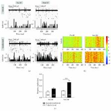
          Mechanisms underlying remifentanil- (RF-) induced hyperalgesia, a phenomenon that is generally named as opioid-induced hyperalgesia (OIH), still remain elusive. The ventral posterior lateral nucleus (VPL) of the thalamus, a key relay station for the transmission of nociceptive information to the cerebral cortex, is activated by RF infusion. Electroacupuncture (EA) is an effective method for the treatment of pain. This study aimed to explore the role of VPL in the development of OIH and the effect of EA treatment on OIH in rats. RF was administered to rats via the tail vein for OIH induction. Paw withdrawal threshold (PWT) in response to mechanical stimuli and paw withdrawal latency (PWL) to thermal stimulation were tested in rats for the assessment of mechanical allodynia and thermal hyperalgesia, respectively. Spontaneous neuronal activity and local field potential (LFP) in VPL were recorded in freely moving rats using the in vivo multichannel recording technique. EA at 2 Hz frequency (pulse width 0.6 ms, 1–3 mA) was applied to the bilateral acupoints “Zusanli” (ST.36) and “Sanyinjiao” (SP.6) in rats. The results showed that both the PWT and PWL were significantly decreased after RF infusion to rats. Meanwhile, both the spontaneous neuronal firing rate and the theta band oscillation in VPL LFP were increased on day 3 post-RF infusion, indicating that the VPL may promote the development of RF-induced hyperalgesia by regulating the pain-related cortical activity. Moreover, 2 Hz-EA reversed the RF-induced decrease both in PWT and PWL of rats and also abrogated the RF-induced augmentation of the spontaneous neuronal activity and the power spectral density (PSD) of the theta band oscillation in VPL LFP. These results suggested that 2 Hz-EA attenuates the remifentanil-induced hyperalgesia via reducing the excitability of VPL neurons and the low-frequency (theta band) oscillation in VPL LFP.

          Related collections

          Most cited references68

          • Record: found
          • Abstract: not found
          • Article: not found

          Ethical guidelines for investigations of experimental pain in conscious animals.

            Bookmark
            • Record: found
            • Abstract: not found
            • Article: not found

            Ethical guidelines for investigations of experimental pain in conscious animals

              Bookmark
              • Record: found
              • Abstract: found
              • Article: not found

              Neural mechanism underlying acupuncture analgesia.

              Acupuncture has been accepted to effectively treat chronic pain by inserting needles into the specific "acupuncture points" (acupoints) on the patient's body. During the last decades, our understanding of how the brain processes acupuncture analgesia has undergone considerable development. Acupuncture analgesia is manifested only when the intricate feeling (soreness, numbness, heaviness and distension) of acupuncture in patients occurs following acupuncture manipulation. Manual acupuncture (MA) is the insertion of an acupuncture needle into acupoint followed by the twisting of the needle up and down by hand. In MA, all types of afferent fibers (Abeta, Adelta and C) are activated. In electrical acupuncture (EA), a stimulating current via the inserted needle is delivered to acupoints. Electrical current intense enough to excite Abeta- and part of Adelta-fibers can induce an analgesic effect. Acupuncture signals ascend mainly through the spinal ventrolateral funiculus to the brain. Many brain nuclei composing a complicated network are involved in processing acupuncture analgesia, including the nucleus raphe magnus (NRM), periaqueductal grey (PAG), locus coeruleus, arcuate nucleus (Arc), preoptic area, nucleus submedius, habenular nucleus, accumbens nucleus, caudate nucleus, septal area, amygdale, etc. Acupuncture analgesia is essentially a manifestation of integrative processes at different levels in the CNS between afferent impulses from pain regions and impulses from acupoints. In the last decade, profound studies on neural mechanisms underlying acupuncture analgesia predominately focus on cellular and molecular substrate and functional brain imaging and have developed rapidly. Diverse signal molecules contribute to mediating acupuncture analgesia, such as opioid peptides (mu-, delta- and kappa-receptors), glutamate (NMDA and AMPA/KA receptors), 5-hydroxytryptamine, and cholecystokinin octapeptide. Among these, the opioid peptides and their receptors in Arc-PAG-NRM-spinal dorsal horn pathway play a pivotal role in mediating acupuncture analgesia. The release of opioid peptides evoked by electroacupuncture is frequency-dependent. EA at 2 and 100Hz produces release of enkephalin and dynorphin in the spinal cord, respectively. CCK-8 antagonizes acupuncture analgesia. The individual differences of acupuncture analgesia are associated with inherited genetic factors and the density of CCK receptors. The brain regions associated with acupuncture analgesia identified in animal experiments were confirmed and further explored in the human brain by means of functional imaging. EA analgesia is likely associated with its counter-regulation to spinal glial activation. PTX-sesntive Gi/o protein- and MAP kinase-mediated signal pathways as well as the downstream events NF-kappaB, c-fos and c-jun play important roles in EA analgesia.
                Bookmark

                Author and article information

                Contributors
                Journal
                Neural Plast
                Neural Plast
                NP
                Neural Plasticity
                Hindawi
                2090-5904
                1687-5443
                2018
                11 November 2018
                : 2018
                : 6109723
                Affiliations
                1Neuroscience Research Institute and Department of Neurobiology, School of Basic Medical Sciences, Peking University, Beijing 100083, China
                2Department of Internal Medicine, Peking University Hospital, Beijing 100871, China
                3The Second Affiliated Hospital of Xinxiang Medical University, Xinxiang, Henan 453002, China
                4Key Laboratory for Neuroscience, Ministry of Education/National Health and Family Planning Commission, Beijing 100083, China
                Author notes

                Academic Editor: Xavier Navarro

                Author information
                http://orcid.org/0000-0002-5454-2759
                http://orcid.org/0000-0002-6473-0585
                http://orcid.org/0000-0003-2506-5137
                http://orcid.org/0000-0001-5670-4857
                Article
                10.1155/2018/6109723
                6252233
                30534151
                d44ea99f-439f-4821-a92e-77cad5a6bdbc
                Copyright © 2018 Hong-Yan Zhao et al.

                This is an open access article distributed under the Creative Commons Attribution License, which permits unrestricted use, distribution, and reproduction in any medium, provided the original work is properly cited.

                History
                : 2 May 2018
                : 4 September 2018
                Funding
                Funded by: China Academy of Chinese Medical Sciences
                Award ID: ZZ04004
                Funded by: the National Basic Research Program of China
                Award ID: 2013CB531905
                Funded by: National Natural Science Foundation of China
                Award ID: 61527815
                Award ID: 81371237
                Award ID: 81671085
                Categories
                Research Article

                Neurosciences
                Neurosciences

                Comments

                Comment on this article