22
views
0
recommends
+1 Recommend
1 collections
    0
    shares
      • Record: found
      • Abstract: found
      • Article: found
      Is Open Access

      Transferrin-targeted porous silicon nanoparticles reduce glioblastoma cell migration across tight extracellular space

      research-article

      Read this article at

      ScienceOpenPublisherPMC
      Bookmark
          There is no author summary for this article yet. Authors can add summaries to their articles on ScienceOpen to make them more accessible to a non-specialist audience.

          Abstract

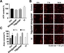
          Mortality of glioblastoma multiforme (GBM) has not improved over the last two decades despite medical breakthroughs in the treatment of other types of cancers. Nanoparticles hold tremendous promise to overcome the pharmacokinetic challenges and off-target adverse effects. However, an inhibitory effect of nanoparticles by themselves on metastasis has not been explored. In this study, we developed transferrin-conjugated porous silicon nanoparticles (Tf@pSiNP) and studied their effect on inhibiting GBM migration by means of a microfluidic-based migration chip. This platform, designed to mimic the tight extracellular migration tracts in brain parenchyma, allowed high-content time-resolved imaging of cell migration. Tf@pSiNP were colloidally stable, biocompatible, and their uptake into GBM cells was enhanced by receptor-mediated internalisation. The migration of Tf@pSiNP-exposed cells across the confined microchannels was suppressed, but unconfined migration was unaffected. The pSiNP-induced destabilisation of focal adhesions at the leading front may partially explain the migration inhibition. More corroborating evidence suggests that pSiNP uptake reduced the plasticity of GBM cells in reducing cell volume, an effect that proved crucial in facilitating migration across the tight confined tracts. We believe that the inhibitory effect of Tf@pSiNP on cell migration, together with the drug-delivery capability of pSiNP, could potentially offer a disruptive strategy to treat GBM.

          Related collections

          Most cited references71

          • Record: found
          • Abstract: found
          • Article: found

          The 2016 World Health Organization Classification of Tumors of the Central Nervous System: a summary.

          The 2016 World Health Organization Classification of Tumors of the Central Nervous System is both a conceptual and practical advance over its 2007 predecessor. For the first time, the WHO classification of CNS tumors uses molecular parameters in addition to histology to define many tumor entities, thus formulating a concept for how CNS tumor diagnoses should be structured in the molecular era. As such, the 2016 CNS WHO presents major restructuring of the diffuse gliomas, medulloblastomas and other embryonal tumors, and incorporates new entities that are defined by both histology and molecular features, including glioblastoma, IDH-wildtype and glioblastoma, IDH-mutant; diffuse midline glioma, H3 K27M-mutant; RELA fusion-positive ependymoma; medulloblastoma, WNT-activated and medulloblastoma, SHH-activated; and embryonal tumour with multilayered rosettes, C19MC-altered. The 2016 edition has added newly recognized neoplasms, and has deleted some entities, variants and patterns that no longer have diagnostic and/or biological relevance. Other notable changes include the addition of brain invasion as a criterion for atypical meningioma and the introduction of a soft tissue-type grading system for the now combined entity of solitary fibrous tumor / hemangiopericytoma-a departure from the manner by which other CNS tumors are graded. Overall, it is hoped that the 2016 CNS WHO will facilitate clinical, experimental and epidemiological studies that will lead to improvements in the lives of patients with brain tumors.
            Bookmark
            • Record: found
            • Abstract: found
            • Article: not found

            Radiotherapy plus Concomitant and Adjuvant Temozolomide for Glioblastoma

            Glioblastoma, the most common primary brain tumor in adults, is usually rapidly fatal. The current standard of care for newly diagnosed glioblastoma is surgical resection to the extent feasible, followed by adjuvant radiotherapy. In this trial we compared radiotherapy alone with radiotherapy plus temozolomide, given concomitantly with and after radiotherapy, in terms of efficacy and safety. Patients with newly diagnosed, histologically confirmed glioblastoma were randomly assigned to receive radiotherapy alone (fractionated focal irradiation in daily fractions of 2 Gy given 5 days per week for 6 weeks, for a total of 60 Gy) or radiotherapy plus continuous daily temozolomide (75 mg per square meter of body-surface area per day, 7 days per week from the first to the last day of radiotherapy), followed by six cycles of adjuvant temozolomide (150 to 200 mg per square meter for 5 days during each 28-day cycle). The primary end point was overall survival. A total of 573 patients from 85 centers underwent randomization. The median age was 56 years, and 84 percent of patients had undergone debulking surgery. At a median follow-up of 28 months, the median survival was 14.6 months with radiotherapy plus temozolomide and 12.1 months with radiotherapy alone. The unadjusted hazard ratio for death in the radiotherapy-plus-temozolomide group was 0.63 (95 percent confidence interval, 0.52 to 0.75; P<0.001 by the log-rank test). The two-year survival rate was 26.5 percent with radiotherapy plus temozolomide and 10.4 percent with radiotherapy alone. Concomitant treatment with radiotherapy plus temozolomide resulted in grade 3 or 4 hematologic toxic effects in 7 percent of patients. The addition of temozolomide to radiotherapy for newly diagnosed glioblastoma resulted in a clinically meaningful and statistically significant survival benefit with minimal additional toxicity. Copyright 2005 Massachusetts Medical Society.
              Bookmark
              • Record: found
              • Abstract: found
              • Article: not found

              The epidemiology of glioma in adults: a "state of the science" review.

              Gliomas are the most common primary intracranial tumor, representing 81% of malignant brain tumors. Although relatively rare, they cause significant mortality and morbidity. Glioblastoma, the most common glioma histology (∼45% of all gliomas), has a 5-year relative survival of ∼5%. A small portion of these tumors are caused by Mendelian disorders, including neurofibromatosis, tuberous sclerosis, and Li-Fraumeni syndrome. Genomic analyses of glioma have also produced new evidence about risk and prognosis. Recently discovered biomarkers that indicate improved survival include O⁶-methylguanine-DNA methyltransferase methylation, isocitrate dehydrogenase mutation, and a glioma cytosine-phosphate-guanine island methylator phenotype. Genome-wide association studies have identified heritable risk alleles within 7 genes that are associated with increased risk of glioma. Many risk factors have been examined as potential contributors to glioma risk. Most significantly, these include an increase in risk by exposure to ionizing radiation and a decrease in risk by history of allergies or atopic disease(s). The potential influence of occupational exposures and cellular phones has also been examined, with inconclusive results. We provide a “state of the science” review of current research into causes and risk factors for gliomas in adults.
                Bookmark

                Author and article information

                Contributors
                nicolas.voelcker@monash.edu
                wingyin.tong@monash.edu
                Journal
                Sci Rep
                Sci Rep
                Scientific Reports
                Nature Publishing Group UK (London )
                2045-2322
                11 February 2020
                11 February 2020
                2020
                : 10
                : 2320
                Affiliations
                [1 ]ISNI 0000000121901201, GRID grid.83440.3b, Department of Pharmaceutical and Biological Chemistry, UCL School of Pharmacy, University College London, Brunswick Square, ; London, United Kingdom
                [2 ]ISNI 0000 0004 1936 7857, GRID grid.1002.3, Drug Delivery, Disposition and Dynamics, Monash Institute of Pharmaceutics Science, Monash University, Parkville, ; Victoria, Australia
                [3 ]ISNI 0000 0004 1937 0482, GRID grid.10784.3a, Department of Biomedical Engineering, The Chinese University of Hong Kong, ; Shatin, New Territories Hong Kong
                [4 ]GRID grid.1016.6, Commonwealth Scientific and Industrial Research Organization (CSIRO), Clayton, ; Victoria, Australia
                [5 ]GRID grid.410660.5, Melbourne Centre for Nanofabrication, Victorian Node of the Australian National Fabrication Facility, Clayton, ; Victoria, Australia
                [6 ]Department of Chemistry, City University of Hong Kong, Kowloon, Hong Kong
                [7 ]ISNI 0000 0001 2097 1371, GRID grid.1374.1, Industrial Physics Laboratory, Department of Physics and Astronomy, University of Turku, ; Turku, Finland
                [8 ]ISNI 0000 0004 1936 7857, GRID grid.1002.3, Department of Materials Science and Engineering, Monash University, Clayton, ; Victoria, Australia
                Author information
                http://orcid.org/0000-0002-1536-7804
                Article
                59146
                10.1038/s41598-020-59146-5
                7012928
                32047170
                d3fc486f-1c1e-4b5e-9798-419373287f3e
                © The Author(s) 2020

                Open Access This article is licensed under a Creative Commons Attribution 4.0 International License, which permits use, sharing, adaptation, distribution and reproduction in any medium or format, as long as you give appropriate credit to the original author(s) and the source, provide a link to the Creative Commons license, and indicate if changes were made. The images or other third party material in this article are included in the article’s Creative Commons license, unless indicated otherwise in a credit line to the material. If material is not included in the article’s Creative Commons license and your intended use is not permitted by statutory regulation or exceeds the permitted use, you will need to obtain permission directly from the copyright holder. To view a copy of this license, visit http://creativecommons.org/licenses/by/4.0/.

                History
                : 17 June 2019
                : 24 January 2020
                Funding
                Funded by: FundRef https://doi.org/10.13039/501100001779, Monash University (MU);
                Funded by: FundRef https://doi.org/10.13039/501100000943, Commonwealth Scientific and Industrial Research Organisation (CSIRO);
                Funded by: FundRef https://doi.org/10.13039/100008716, Science and Industry Endowment Fund (Science & Industry Endowment Fund);
                Award ID: PF16-072
                Award Recipient :
                Categories
                Article
                Custom metadata
                © The Author(s) 2020

                Uncategorized
                biomaterials,cns cancer
                Uncategorized
                biomaterials, cns cancer

                Comments

                Comment on this article