213
views
0
recommends
+1 Recommend
1 collections
    0
    shares
      • Record: found
      • Abstract: found
      • Article: found

      Cellular and molecular mechanisms of mitochondrial function

      review-article

      Read this article at

      ScienceOpenPublisherPMC
      Bookmark
          There is no author summary for this article yet. Authors can add summaries to their articles on ScienceOpen to make them more accessible to a non-specialist audience.

          Abstract

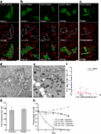
          Mitochondria are membrane bound organelles present in almost all eukaryotic cells. Responsible for orchestrating cellular energy production, they are central to the maintenance of life and the gatekeepers of cell death. Thought to have originated from symbiotic ancestors, they carry a residual genome as mtDNA encoding 13 proteins essential for respiratory chain function. Mitochondria comprise an inner and outer membrane that separate and maintain the aqueous regions, the intermembrane space and the matrix. Mitochondria contribute to many processes central to cellular function and dysfunction including calcium signalling, cell growth and differentiation, cell cycle control and cell death. Mitochondrial shape and positioning in cells is crucial and is tightly regulated by processes of fission and fusion, biogenesis and autophagy, ensuring a relatively constant mitochondrial population. Mitochondrial dysfunction is implicated in metabolic and age related disorders, neurodegenerative diseases and ischemic injury in heart and brain.

          Related collections

          Most cited references89

          • Record: found
          • Abstract: found
          • Article: not found

          Sequence and organization of the human mitochondrial genome.

          The complete sequence of the 16,569-base pair human mitochondrial genome is presented. The genes for the 12S and 16S rRNAs, 22 tRNAs, cytochrome c oxidase subunits I, II and III, ATPase subunit 6, cytochrome b and eight other predicted protein coding genes have been located. The sequence shows extreme economy in that the genes have none or only a few noncoding bases between them, and in many cases the termination codons are not coded in the DNA but are created post-transcriptionally by polyadenylation of the mRNAs.
            Bookmark
            • Record: found
            • Abstract: found
            • Article: found
            Is Open Access

            Parkin is recruited selectively to impaired mitochondria and promotes their autophagy

            Loss-of-function mutations in Park2, the gene coding for the ubiquitin ligase Parkin, are a significant cause of early onset Parkinson's disease. Although the role of Parkin in neuron maintenance is unknown, recent work has linked Parkin to the regulation of mitochondria. Its loss is associated with swollen mitochondria and muscle degeneration in Drosophila melanogaster, as well as mitochondrial dysfunction and increased susceptibility to mitochondrial toxins in other species. Here, we show that Parkin is selectively recruited to dysfunctional mitochondria with low membrane potential in mammalian cells. After recruitment, Parkin mediates the engulfment of mitochondria by autophagosomes and the selective elimination of impaired mitochondria. These results show that Parkin promotes autophagy of damaged mitochondria and implicate a failure to eliminate dysfunctional mitochondria in the pathogenesis of Parkinson's disease.
              Bookmark
              • Record: found
              • Abstract: found
              • Article: not found

              Molecular characterization of mitochondrial apoptosis-inducing factor.

              Mitochondria play a key part in the regulation of apoptosis (cell death). Their intermembrane space contains several proteins that are liberated through the outer membrane in order to participate in the degradation phase of apoptosis. Here we report the identification and cloning of an apoptosis-inducing factor, AIF, which is sufficient to induce apoptosis of isolated nuclei. AIF is a flavoprotein of relative molecular mass 57,000 which shares homology with the bacterial oxidoreductases; it is normally confined to mitochondria but translocates to the nucleus when apoptosis is induced. Recombinant AIF causes chromatin condensation in isolated nuclei and large-scale fragmentation of DNA. It induces purified mitochondria to release the apoptogenic proteins cytochrome c and caspase-9. Microinjection of AIF into the cytoplasm of intact cells induces condensation of chromatin, dissipation of the mitochondrial transmembrane potential, and exposure of phosphatidylserine in the plasma membrane. None of these effects is prevented by the wide-ranging caspase inhibitor known as Z-VAD.fmk. Overexpression of Bcl-2, which controls the opening of mitochondrial permeability transition pores, prevents the release of AIF from the mitochondrion but does not affect its apoptogenic activity. These results indicate that AIF is a mitochondrial effector of apoptotic cell death.
                Bookmark

                Author and article information

                Journal
                Best Pract Res Clin Endocrinol Metab
                Best Pract. Res. Clin. Endocrinol. Metab
                Best Practice & Research. Clinical Endocrinology & Metabolism
                Elsevier
                1521-690X
                1878-1594
                December 2012
                December 2012
                : 26
                : 6
                : 711-723
                Affiliations
                [a ]Department of Cell and Developmental Biology, University College London, London WC1E 6BT, United Kingdom
                [b ]UK Parkinson’s Disease Consortium, Institute of Neurology, University College London, London WC1N 3BG, United Kingdom
                [c ]Department of Physics and Astronomy, University College London, London WC1E 6BT, United Kingdom
                Author notes
                []Corresponding author. Department of Cell and Developmental Biology, University College London, London WC1E 6BT, United Kingdom. Tel.: +44 20 7679 3207. m.duchen@ 123456ucl.ac.uk
                [d]

                Tel.: +44 20 7679 7127.

                Article
                YBEEM834
                10.1016/j.beem.2012.05.003
                3513836
                23168274
                d2d11532-8394-4d43-a16e-e777effab52e
                © 2012 Elsevier Ltd.

                This document may be redistributed and reused, subject to certain conditions.

                History
                Categories
                1

                Endocrinology & Diabetes
                mitochondria,oxidative phosphorylation,apoptosis,intracellular calcium,mitochondrial fission

                Comments

                Comment on this article

                scite_
                0
                0
                0
                0
                Smart Citations
                0
                0
                0
                0
                Citing PublicationsSupportingMentioningContrasting
                View Citations

                See how this article has been cited at scite.ai

                scite shows how a scientific paper has been cited by providing the context of the citation, a classification describing whether it supports, mentions, or contrasts the cited claim, and a label indicating in which section the citation was made.

                Similar content536

                Cited by282

                Most referenced authors2,202