5
views
0
recommends
+1 Recommend
0 collections
    0
    shares
      • Record: found
      • Abstract: found
      • Article: found
      Is Open Access

      DTI-ALPS: An MR biomarker for motor dysfunction in patients with subacute ischemic stroke

      research-article

      Read this article at

      Bookmark
          There is no author summary for this article yet. Authors can add summaries to their articles on ScienceOpen to make them more accessible to a non-specialist audience.

          Abstract

          Purpose

          Brain glymphatic dysfunction is involved in the pathologic process of acute ischemic stroke (IS). The relationship between brain glymphatic activity and dysfunction in subacute IS has not been fully elucidated. Diffusion tensor image analysis along the perivascular space (DTI-ALPS) index was used in this study to explore whether glymphatic activity was related to motor dysfunction in subacute IS patients.

          Methods

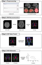
          Twenty-six subacute IS patients with a single lesion in the left subcortical region and 32 healthy controls (HCs) were recruited in this study. The DTI-ALPS index and DTI metrics (fractional anisotropy, FA, and mean diffusivity, MD) were compared within and between groups. Spearman's and Pearson's partial correlation analyses were performed to analyze the relationships of the DTI-ALPS index with Fugl-Meyer assessment (FMA) scores and with corticospinal tract (CST) integrity in the IS group, respectively.

          Results

          Six IS patients and two HCs were excluded. The left DTI-ALPS index of the IS group was significantly lower than that of the HC group ( t = −3.02, p = 0.004). In the IS group, a positive correlation between the left DTI-ALPS index and the simple Fugl-Meyer motor function score (ρ = 0.52, p = 0.019) and a significant negative correlation between the left DTI-ALPS index and the FA ( R = −0.55, p = 0.023) and MD ( R = −0.48, p = 0.032) values of the right CST were found.

          Conclusions

          Glymphatic dysfunction is involved in subacute IS. DTI-ALPS could be a potential magnetic resonance (MR) biomarker of motor dysfunction in subacute IS patients. These findings contribute to a better understanding of the pathophysiological mechanisms of IS and provide a new target for alternative treatments for IS.

          Related collections

          Most cited references101

          • Record: found
          • Abstract: found
          • Article: not found

          Fast robust automated brain extraction.

          An automated method for segmenting magnetic resonance head images into brain and non-brain has been developed. It is very robust and accurate and has been tested on thousands of data sets from a wide variety of scanners and taken with a wide variety of MR sequences. The method, Brain Extraction Tool (BET), uses a deformable model that evolves to fit the brain's surface by the application of a set of locally adaptive model forces. The method is very fast and requires no preregistration or other pre-processing before being applied. We describe the new method and give examples of results and the results of extensive quantitative testing against "gold-standard" hand segmentations, and two other popular automated methods. Copyright 2002 Wiley-Liss, Inc.
            Bookmark
            • Record: found
            • Abstract: found
            • Article: not found

            A paravascular pathway facilitates CSF flow through the brain parenchyma and the clearance of interstitial solutes, including amyloid β.

            Because it lacks a lymphatic circulation, the brain must clear extracellular proteins by an alternative mechanism. The cerebrospinal fluid (CSF) functions as a sink for brain extracellular solutes, but it is not clear how solutes from the brain interstitium move from the parenchyma to the CSF. We demonstrate that a substantial portion of subarachnoid CSF cycles through the brain interstitial space. On the basis of in vivo two-photon imaging of small fluorescent tracers, we showed that CSF enters the parenchyma along paravascular spaces that surround penetrating arteries and that brain interstitial fluid is cleared along paravenous drainage pathways. Animals lacking the water channel aquaporin-4 (AQP4) in astrocytes exhibit slowed CSF influx through this system and a ~70% reduction in interstitial solute clearance, suggesting that the bulk fluid flow between these anatomical influx and efflux routes is supported by astrocytic water transport. Fluorescent-tagged amyloid β, a peptide thought to be pathogenic in Alzheimer's disease, was transported along this route, and deletion of the Aqp4 gene suppressed the clearance of soluble amyloid β, suggesting that this pathway may remove amyloid β from the central nervous system. Clearance through paravenous flow may also regulate extracellular levels of proteins involved with neurodegenerative conditions, its impairment perhaps contributing to the mis-accumulation of soluble proteins.
              Bookmark
              • Record: found
              • Abstract: found
              • Article: not found

              Structural and functional features of central nervous system lymphatics

              One of the characteristics of the CNS is the lack of a classical lymphatic drainage system. Although it is now accepted that the CNS undergoes constant immune surveillance that takes place within the meningeal compartment 1–3 , the mechanisms governing the entrance and exit of immune cells from the CNS remain poorly understood 4–6 . In searching for T cell gateways into and out of the meninges, we discovered functional lymphatic vessels lining the dural sinuses. These structures express all of the molecular hallmarks of lymphatic endothelial cells, are able to carry both fluid and immune cells from the CSF, and are connected to the deep cervical lymph nodes. The unique location of these vessels may have impeded their discovery to date, thereby contributing to the long-held concept of the absence of lymphatic vasculature in the CNS. The discovery of the CNS lymphatic system may call for a reassessment of basic assumptions in neuroimmunology and shed new light on the etiology of neuroinflammatory and neurodegenerative diseases associated with immune system dysfunction.
                Bookmark

                Author and article information

                Contributors
                Journal
                Front Neurosci
                Front Neurosci
                Front. Neurosci.
                Frontiers in Neuroscience
                Frontiers Media S.A.
                1662-4548
                1662-453X
                31 March 2023
                2023
                : 17
                : 1132393
                Affiliations
                [1] 1Department of Medical Imaging, The First Affiliated Hospital of Xi'an Jiaotong University , Xi'an, China
                [2] 2Department of Radiology, Xi'an Daxing Hospital , Xi'an, China
                [3] 3Department of Rehabilitation Medicine, Xi'an Daxing Hospital , Xi'an, China
                Author notes

                Edited by: Shugeng Chen, Fudan University, China

                Reviewed by: Toshiaki Taoka, Nagoya University, Japan; Hongbin Han, Peking University Third Hospital, China; Jesse Klostranec, McGill University Health Centre, Canada

                *Correspondence: Ming Zhang zmdx_2022@ 123456163.com

                This article was submitted to Neural Technology, a section of the journal Frontiers in Neuroscience

                †These authors have contributed equally to this work

                Article
                10.3389/fnins.2023.1132393
                10102345
                37065921
                d2aad5ab-442f-46d4-b9c2-ab4da6367ee2
                Copyright © 2023 Qin, Li, Qiao, Zou, Qian, Li, Zhu, Huo, Wang and Zhang.

                This is an open-access article distributed under the terms of the Creative Commons Attribution License (CC BY). The use, distribution or reproduction in other forums is permitted, provided the original author(s) and the copyright owner(s) are credited and that the original publication in this journal is cited, in accordance with accepted academic practice. No use, distribution or reproduction is permitted which does not comply with these terms.

                History
                : 27 December 2022
                : 03 March 2023
                Page count
                Figures: 6, Tables: 1, Equations: 1, References: 101, Pages: 12, Words: 9126
                Categories
                Neuroscience
                Original Research

                Neurosciences
                glymphatic system,ischemic stroke,analysis along perivascular space,corticospinal tract,motor dysfunction

                Comments

                Comment on this article