51
views
0
recommends
+1 Recommend
1 collections
    1
    shares

      Authors - did you know SICOT-J has been awarded the DOAJ Seal for "best practice in open access publishing"?

      • Indexed in Scopus
      • 30% discount on article processing charges (APCs) for members of SICOT

      Instructions for authors, online submissions and free e-mail alerts all available here 

      • Record: found
      • Abstract: found
      • Article: found
      Is Open Access

      Intraosseous schwannoma of the glenoid: case report and literature review

      research-article
      1 , * , 1 , 1 , 1 , 1
      SICOT-J
      EDP Sciences
      Schwannoma, Neurilemmoma, Intraosseous, Glenoid

      Read this article at

      ScienceOpenPublisherPMC
      Bookmark
          There is no author summary for this article yet. Authors can add summaries to their articles on ScienceOpen to make them more accessible to a non-specialist audience.

          Abstract

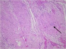
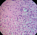
          Intraosseous schwannomas represent an extremely rare subgroup of schwannomas, accounting for <1% of all primary bone tumors. They mostly occur in the mandible, the maxilla, the sacrum, and they are also seen in long bones. We herein report a rare presentation of an intraosseous schwannoma in the glenoid of a 49-year-old patient. She complained of shoulder pain and was referred to the orthopaedic oncologist after detection of a suspicious lesion on imaging. Biopsy revealed benign spindle cells and immunohistochemistry was positive for S100. Because of the rarity of these intraosseous schwannomas it is important to recognize their radiological and histological features and make a differential diagnosis with other lytic tumors. Only if these characteristics are recognized, correct treatment can be given with definite curettage and bone grafting and correct follow-up with avoidance of unnecessary adjuvant therapy.

          Related collections

          Most cited references22

          • Record: found
          • Abstract: found
          • Article: not found

          Spinal dumbbell tumors: an analysis of a series of 118 cases.

          The authors analyzed a series of 118 cases of spinal dumbbell tumors to elucidate the feature of the tumors. Of 674 cases of spinal cord tumors, the incidence of dumbbell tumors was studied. The tumors were analyzed, and the authors focus on the distribution of age and sex, the pathological diagnoses, their locations, Eden classification, and the surgical methods used. The incidence of dumbbell tumors was 18%. The mean patient age was 43 years, which was younger than that for all spinal cord tumors (mean 50 years). There were 11 patients younger than 10 years of age. The rate of dumbbell tumors in the cervical spine was significantly higher than that of all spinal cord tumors. Fifteen (18%) of the 81 schwannomas were observed in the C-2 nerve root, thus having a higher incidence than those in the other nerve roots. In 99 cases (84%), the tumors were removed through a hemilaminectomy with or without a facetectomy and posterior fusion. Of 118 cases, 69% of the tumors were schwannomas, and malignant tumors were found in 10 cases (8.5%). Seven (64%) of 11 patients younger than 10 years of age had malignant tumors. Three patients older than 10 years of age had malignant tumors, thus accounting for 2.8% of the 107 older patients. The incidence of dumbbell tumors was 18%, and they are not uncommon. Malignant dumbbell tumors were more common in children younger than 10 years of age than in older patients.
            Bookmark
            • Record: found
            • Abstract: found
            • Article: found
            Is Open Access

            Learning from eponyms: Jose Verocay and Verocay bodies, Antoni A and B areas, Nils Antoni and Schwannomas

            Schwannomas are benign peripheral nerve sheath neoplasms composed almost entirely of Schwann cells and are diagnosed histopathologically by the presence of singular architectural patterns called Antoni A and Antoni B areas. These were described first in 1920 by the Swedish neurologist Nils Antoni. The Antoni A tissue is highly cellular and made up of palisades of Schwann cell nuclei, a pattern first described in 1910 by the Uruguayan neuro-pathologist Jose Verocay and are known as Verocay bodies. This article describes the structure and appearance of Verocay bodies and Antoni A and B areas with a brief biographical introduction of the men who described these patterns.
              Bookmark
              • Record: found
              • Abstract: found
              • Article: not found

              Peripheral Nerve Schwannoma: A Review of Varying Clinical Presentations and Imaging Findings.

              A schwannoma or neurilemmoma is a benign, isolated, noninvasive, and encapsulated tumor originating from Schwann cells of the peripheral nerve sheath. The incidence of a schwannoma occurring in the foot and ankle is rare, with prevalence rate of 1% to 10%. Schwannomas have no sex predilection, and they commonly occur in patients in their fourth decade. Malignant transformation of benign schwannoma is unusual; however, it is important to note that malignant variants of schwannomas do exist and account for about 5% to 10% of all soft tissue sarcomas. We present 3 cases of benign schwannoma in the lower extremity. All 3 patients presented with varying clinical symptoms, including pain, paresthesia, weakness, and a palpable mass. A schwannoma was eventually diagnosed in all 3 patients. We discuss and review the known entities of peripheral nerve schwannoma and describe the clinical and imaging findings and therapeutic strategies for treating and diagnosing peripheral nerve schwannoma.
                Bookmark

                Author and article information

                Journal
                SICOT J
                SICOT J
                sicotj
                SICOT-J
                EDP Sciences
                2426-8887
                2021
                08 January 2021
                : 7
                : ( publisher-idID: sicotj/2021/01 )
                : 2
                Affiliations
                [1 ] University Hospitals Leuven Campus Gasthuisberg Herestraat 49 3000 Leuven Belgium
                Author notes
                [* ]Corresponding author: preyniers@ 123456gmail.com
                Author information
                http://orcid.org/0000-0001-7098-7486
                http://orcid.org/0000-0003-4321-7771
                http://orcid.org/0000-0003-2244-5839
                Article
                sicotj200083 10.1051/sicotj/2020048
                10.1051/sicotj/2020048
                7792494
                33416488
                d2871dd8-1656-4df5-87aa-3c86ebccc589
                © The Authors, published by EDP Sciences, 2021

                This is an Open Access article distributed under the terms of the Creative Commons Attribution License ( https://creativecommons.org/licenses/by/4.0), which permits unrestricted use, distribution, and reproduction in any medium, provided the original work is properly cited.

                History
                : 06 July 2020
                : 13 December 2020
                Page count
                Figures: 2, Tables: 0, Equations: 0, References: 19, Pages: 4
                Categories
                Shoulder
                Original Article

                schwannoma,neurilemmoma,intraosseous,glenoid
                schwannoma, neurilemmoma, intraosseous, glenoid

                Comments

                Comment on this article